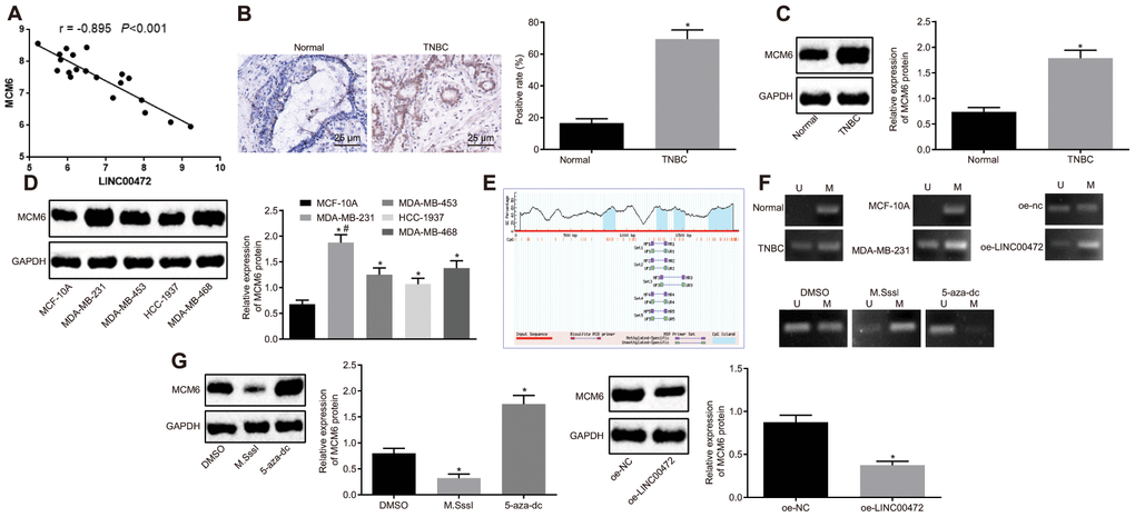
Figure 3.LINC00472 increases MCM6 promoter methylation to inhibit the MCM6 expression. (A) A negative correlation between LINC00472 and MCM6 in the microarray dataset GSE61724. (B) The expression of MCM6 in TNBC tissues normalized to GAPDH identified by means of immunohistochemistry (× 400), * p < 0.05 vs. the normal tissues. (C) The expression of MCM6 in TNBC tissues normalized to GAPDH as determined by means of Western blot analysis, * p < 0.05 vs. the normal tissues. (D) The expression of MCM6 in TNBC cell lines normalized to GAPDH determined by means of Western blot analysis, * p < 0.05 vs. the MCF-10A cell line, # p < 0.05 vs. the HCC-1937 or MDA-MB-468 cell line. (E) The CpG island in the MCM6 promoter region predicted on MethPrimer website (https://www.urogene.org). (F) The methylation in the MCM6 promoter region as detected by means of MSP assay. (G) The expression of MCM6 in MDA-MB-231 cells normalized to GAPDH after treatment with M.SssI or 5-aza-dc or after transfection with oe-LINC00472 determined by means of Western blot analysis, * p < 0.05 vs. the DMSO (MDA-MB-231 cells treated with DMSO) or oe-NC group (MDA-MB-231 cells transfected with oe-NC). The results were measurement data and expressed as mean ± standard deviation. Data comparison in (B and C) was analyzed by the paired t-test, in (D) was analyzed by one-way ANOVA followed by Tukey’s post hoc test and in (G) was analyzed by the unpaired t-test. The experiment was conducted 3 times independently.