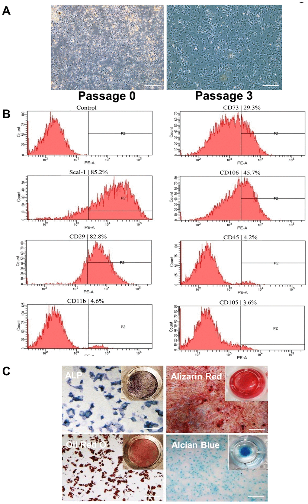
Figure 1.Isolation and identification of BMSCs by phenotypic characterization and multipotent differentiation potential. (A) Cells were isolated from the femurs and tibias of 3- to 4-week-old mice shown at P0 and P3. Cells are attached at P3. Scale Bar=200μm. (B) Flow cytometric analysis of cell surface markers on isolated BMSCs indicates Scal-1+ CD29+ CD11b- CD45- CD105-. (C) Differentiation capacity of BMSCs: ALP staining of cells cultured in osteogenic induction medium for 7 days (upper-left image); alizarin red staining of cells cultured in osteogenic induction medium for 21 days (upper-right image); oil red O staining of cells cultured in adipogenic induction medium for 7 days (lower-left image); and alcian blue staining of cells cultured in chondrogenic induction medium for 14 days (lower-right image). P0, passage 0; P2, passage 2; P3, passage 3. Scale Bar=200μm.