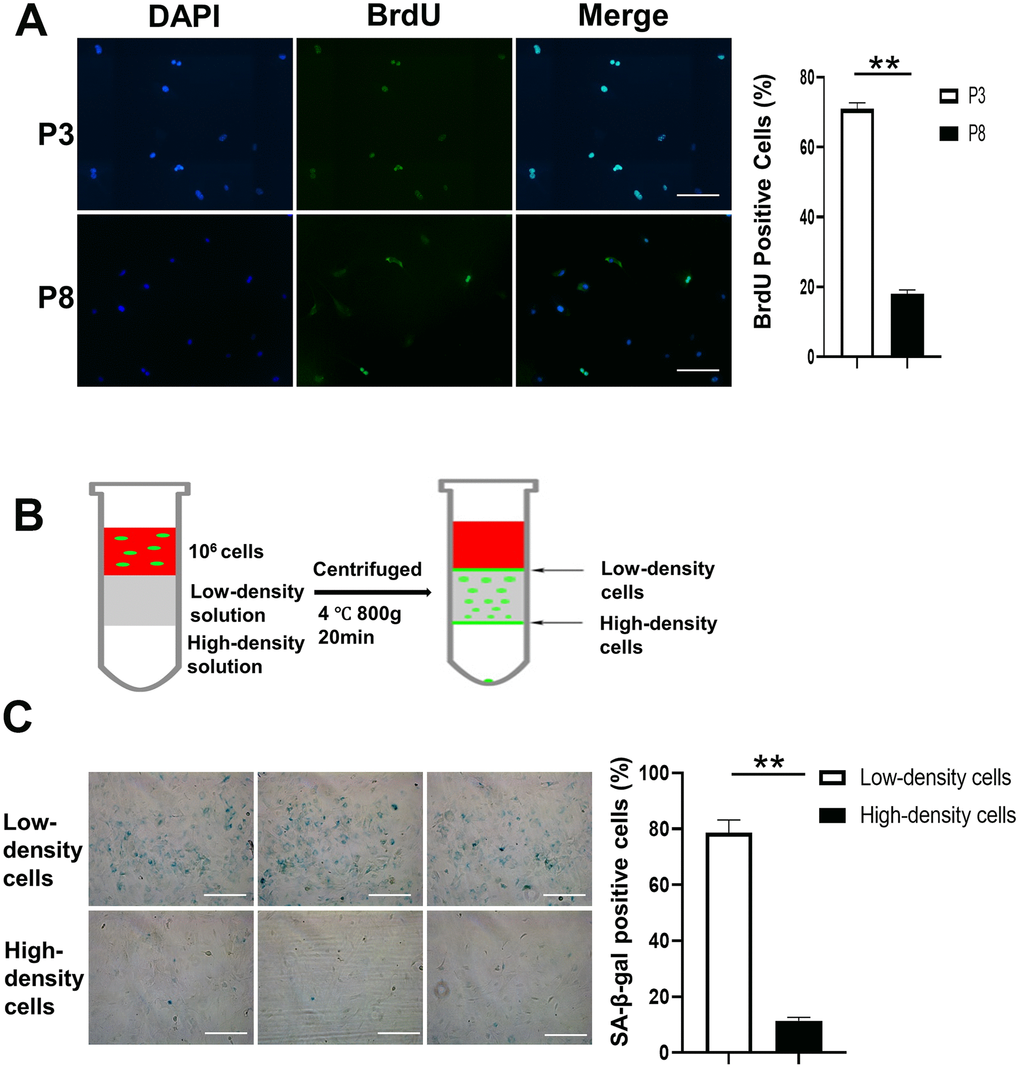
Figure 2.Density gradient separation of proliferating and senescent cells. (A) Immunofluorescence of BrdU positive cells is shown in the first panel. The right panel shows that the number of BrdU positive cells is significantly lower in P8 compared to P3. Scale Bar=200μm. (B) BMSCs (P8), a mixture of proliferating and senescent cells, were centrifuged through a density gradient medium (OptiPrep, Sigma-Aldrich) at 800g for 20 minutes. Aliquots (0.5 mL) were taken from the low- and high-density layers. The cells were then incubated in a 48-well plate. (C) SA-β-gal staining of the 2 groups. Low-density cells contained a higher percentage of SA-β-gal positive cells compared to the high-density cells. The right panel shows that the number of SA-β-gal positive cells is significantly lower in high-density cells compared to low-density cells (n=3). BrdU indicates bromodeoxyuridine; DAPI, 4′,6-diamidino-2-phenylindole; P3, passage 3; P8, passage 8; SA-β-gal, senescence-associated β-galactosidase; Paired T-Test **, P<.01. data are represented as mean ± SEM. Scale Bar=200μm.