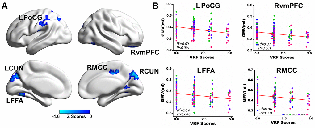
Figure 2.Multivariate regression analysis indicates the effects of VRFs on gray matter volume across all subjects. (A) Brain regions representing the significant effects of VRFs on GMV after controlling for the effects of covariates, including age, gender, APOEε4 genotype, and group. The blue color indicates a negative correlation between VRF scores and GMV. The color bar is presented with z scores. (B) Representative illustration of the significant effects of VRFs on regions of the LPoCG, LFFA, RvmPFC and RMCC. Abbreviations: VRFs, vascular risk factors; LPoCG, left postcentral gyrus; CUN, cuneus; LFFA, left fusiform face area; RvmPFC, right ventromedial prefrontal cortex; RMCC, right middle cingulate cortex; GMV, gray matter volume; CN, cognitively normal; EMCI, early mild cognitive impairment; LMCI, late mild cognitive impairment; AD, Alzheimer’s disease; APOE, apolipoprotein E.