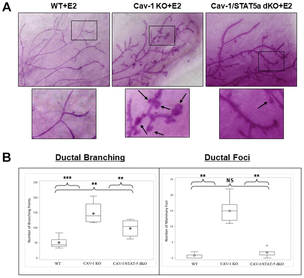
Figure 2.Deletion of STAT5a in Cav-1 KO mice prevents mammary branching and DCIS-Like Foci formation. (A) Mammary glands of estrogen-treated WT, Cav-1 KO, and Cav-1/STAT5a dKO mice were subjected to whole mount analysis to assess ductal branching and foci formation. Images of the mammary gland whole mounts were captured at 40x objective using an Olympus DP71 camera. Black arrows indicate mammary foci. Original cohort sizes were as follows: WT+E2 (9 mice); Cav-1 KO+E2 (7 mice); Cav-1/STAT5a dKO+E2 (8 mice). (B) SAS programming software (version 9.4) was used to generate box plots displaying the number of ductal branching points (left panel) and the number of ductal foci (right panel) for each experimental group. Ductal branching was calculated as a summation of primary, secondary, and tertiary branch points. The absence of STAT5a in the Cav-1 KO mammary gland led to a decrease in both ductal branching and foci formation. Quantitatively, changes in ductal branching were as follows (left panel): WT vs. Cav-1 KO (2.9-fold, p<0.001), Cav-1 KO vs. Cav-1/STAT5a dKO (1.5-fold, p<0.01), WT vs. Cav-1/STAT5a dKO (1.9-fold, p<0.01). Quantitatively, changes in ductal foci were as follows (right panel): WT vs. Cav-1 KO (19.3-fold, p<0.01), Cav-1 KO vs. Cav-1/STAT5a dKO (8.6-fold, p<0.01), WT vs. Cav-1/STAT5a dKO (NS, p=0.164).