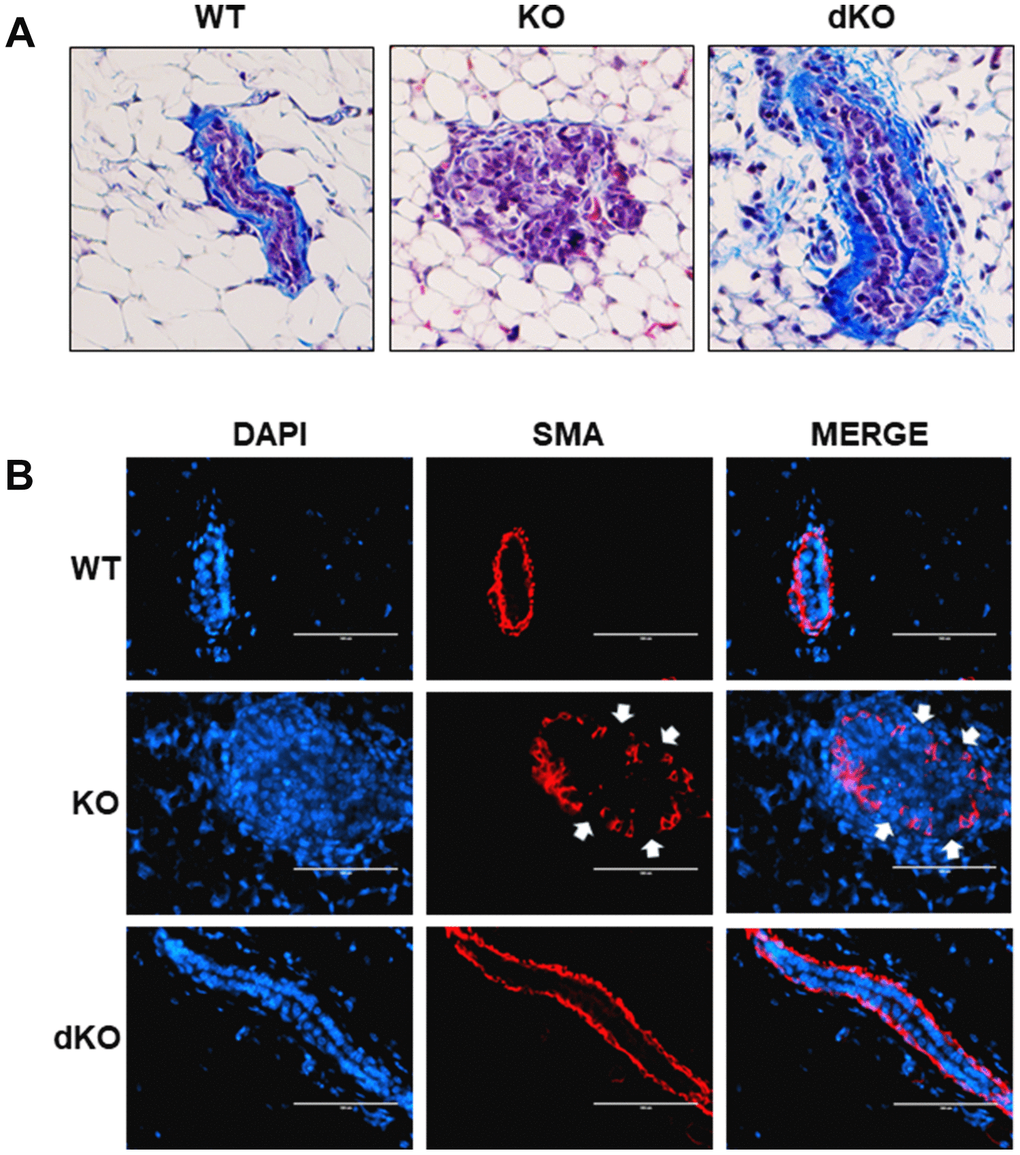
Figure 4.Collagen and smooth muscle actin layer remain uninterrupted in Cav-1/STAT5a dKO mice mammary ducts following estrogen treatment. (A) Mammary glands of estrogen-treated WT, Cav-1 KO, and Cav-1/STAT5a dKO mice were stained with Masson’s trichrome to highlight the collagen (blue staining) lining the outside of the basement membrane of the ducts. Qualitatively, Cav-1 KO mammary glands stimulated with estrogen demonstrated a complete degradation of collagen surrounding the basement membrane, whereas WT ducts showed intact collagen deposition. Deletion of STAT5a from estrogen-treated Cav-1 KO mice restored collagen deposition to WT levels. For each experimental group, trichrome staining was performed in triplicate on mammary glands derived from 3 independent mice. (B) Mammary glands of estrogen-treated WT, Cav-1 KO, and Cav-1/STAT5a dKO mice were immunostained with an antibody recognizing alpha smooth muscle actin (SMA) to highlight the myoepithelial cells lining the inside of the basement membrane of the ducts. DAPI was used as a nuclear counterstain. The EVOS FL microscope was used to capture images at 40x objective with the DAPI and Texas Red light cubes (blue: DAPI immunostaining; red: SMA immunostaining). Qualitatively, the SMA layer surrounding estrogen-treated Cav-1 KO mammary ducts was disrupted (white arrows indicate breaks in the myoepithelial cells), but completely intact around WT ducts. Cav-1 KO mice lacking STAT5a expression maintained an intact SMA layer similar to WT mice. For each experimental group, immunofluorescence was performed in triplicate on mammary glands derived from 3 independent mice.