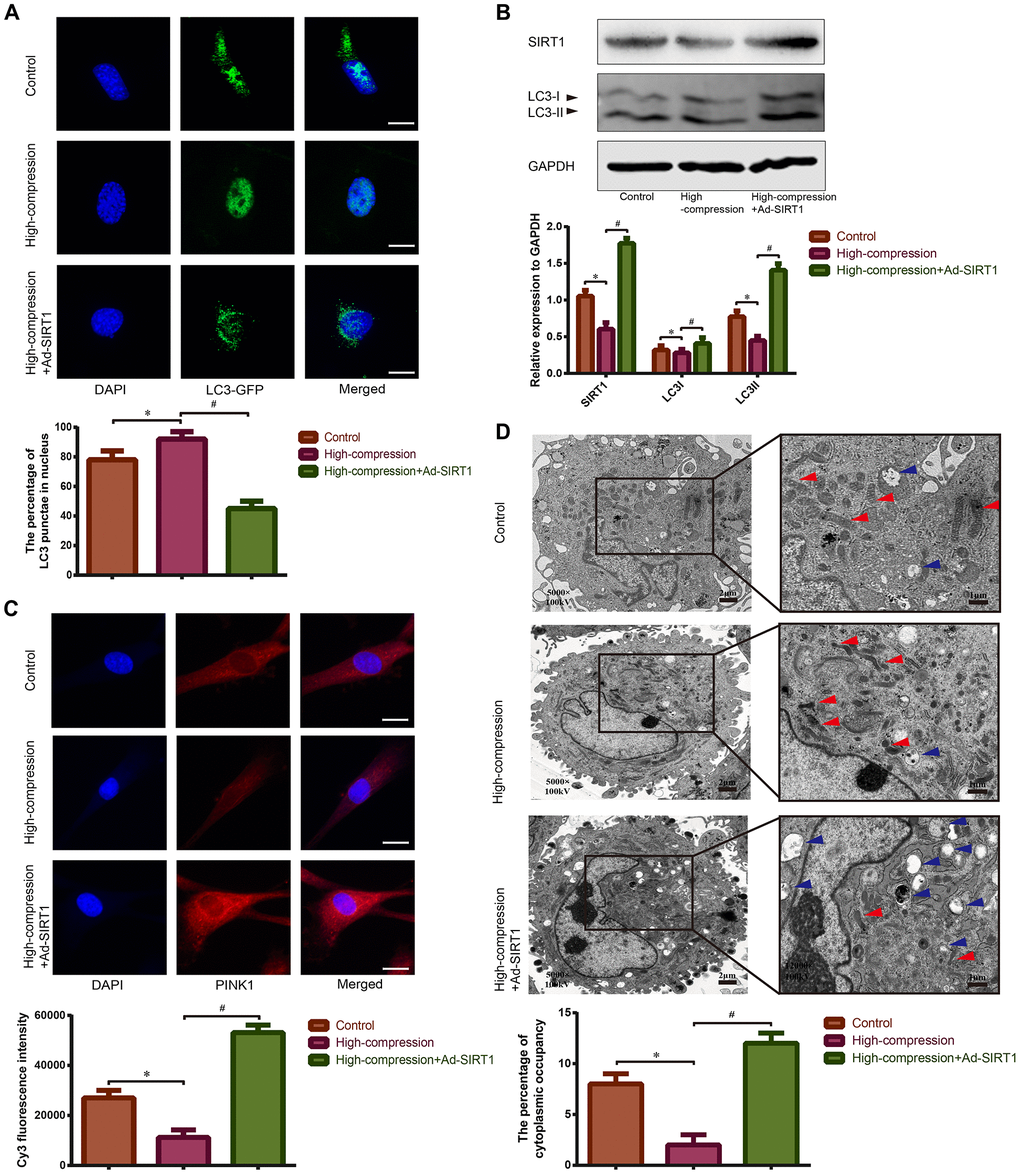
Figure 4.Observation of mitophagy in human NP cells with or without Ad-SIRT1 treatment under high-magnitude compression. (A) The distribution of GFP-LC3 puncta in NP cells subjected to high-compression or high-compression plus Ad-SIRT1 treatment was visualized by confocal microscopy (400×) after transfection of GFP-LC3-expressing adenovirus. (B) The relative expression levels of SIRT1 and LC3II/I in NP cells subject to high-compression or high-compression plus Ad-SIRT1 treatment were analyzed by Western blotting. (C) The expression of the key mitophagic regulator PINK1 was observed and analyzed by fluorescence microscopy (400×) after immunofluorescence staining. (D) The electron micrographs of mitochondria and autophagosomes in NP cells were observed using TEM (5000×), and double-membrane profiles resembling pieces of mitochondria could be found in some autophagosomes in the Ad-SIRT1-treated group. Blue arrows show the typical autophagic vacuole double-layer ultrastructural morphology. Red arrows represent the electron micrographs of mitochondria in the NP cells from each group. *P<0.05 versus the control group. #P<0.05 versus the high-compression group. White bars=100 μm. Black bars=2 μm.