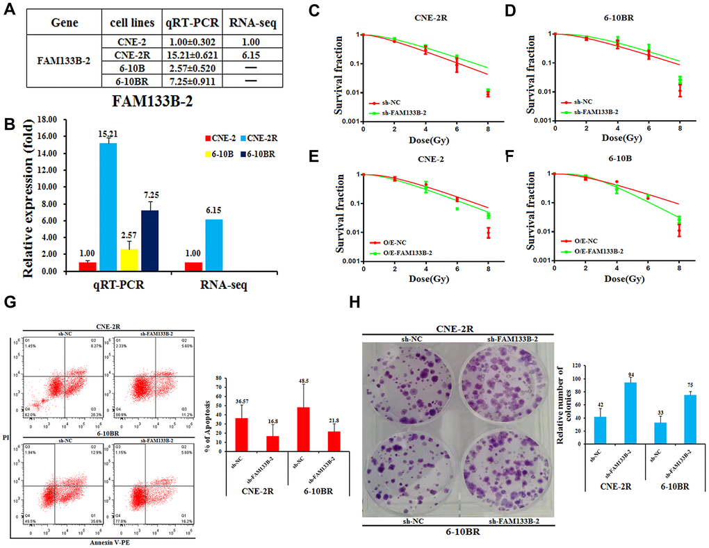
Figure 2.Effects of a forced reversal of FAM133B-2 level on the nasopharyngeal cancer cells. The relative FAM133B-2 level (fold) in CNE-2R and 6-10BR cells versus CNE-2 and 6-10B cells measured by both miR-omic and qRT-PCR analyses is shown in a table (A) and those measured by qRT-PCR are shown in a plot (B). “-” indicates no detection in the omic analysis. sh-FAM133B-2-transfected CNE-2R (C) and 6-10BR (D) cells survival fraction versus the negative control (NC) cells for 24h, then cells were digested and counted according to 500 (0Gy), 1000 (2Gy), 2000 (4Gy), 5000 (6Gy), 8000 (8Gy) cells/well and was inoculated in a 6-well plate in triplicate, the corresponding dose was irradiated after 24h, using a 6-MV x-ray generated by a linear accelerator Varian trilogy at a dose rate of 2Gy/min (Varian trilogy at a dose rate of 2Gy/min). CNE-2 (E) and 6-10B (F) cells infected with FAM133B-2-O/E versus the negative control (NC-O/E), then were digested and counted according to 500 (0Gy), 1000 (2Gy), 2000 (4Gy), 5000 (6Gy), 8000 (8Gy) cells/well and was inoculated in a 6-well plate in triplicate, the corresponding dose was irradiated after 24h, using a 6-MV x-ray generated by a linear accelerator Varian trilogy at a dose rate of 2Gy/min (Varian trilogy at a dose rate of 2Gy/min). (G) The effects of the forced reversal of FAM133B-2 level on the apoptosis of CNE-2R and 6-10BR cells by FACS analysis in plot and in the original with a graph of the analyzed data and plots of the original FACS data. (H) The effects of the forced reversal of FAM133B-2 level on the sphere numbers of CNE-2Rand 6-10BR cells. The sphere numbers were determined after seven days for the first generation (G1) and seven days after seeding for G2. Treatment with SCF was repeated when the cells were passaged. Colony formation numbers, relative sphere formation are shown. The sphere formation assays showed that the sphere numbers of CNE-2R and 6-10BR cells was fewer than that of parental cells. The data are mean±SD of two independent experiments. The surviving fraction was calculated using the multitarget single-hit model: Y=1-(1-exp(-k*x))^N. The data are presented as the mean±standard deviation of results from 3 independent experiments, and two way Anova was used to calculate statistical significance.