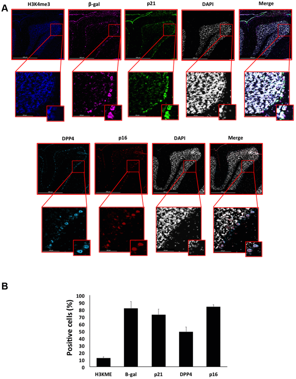
Figure 7.(A) Immunofluorescent staining of senescence markers in Purkinje cells of 9 months old C57/Bl6J mouse cerebellar cortex. Images display H3K4me3 (blue), β-gal (purple), p21 (green), DPP4 (cyan) p16 (red) and DAPI-stained nuclei (white) fluorescence signals. The corresponding multichannel overlaid images are shown in the right column. All these markers, except H3K4me3, show an increased localization and expression in Purkinje cells. Representative images from 3 different mice are shown. (B) Frequency of positive cells for each marker as in (A), indicated in %.