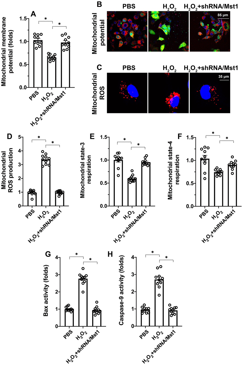
Figure 2.Mst1 induces mitochondrial dysfunction in oxidative stress-induced RA-FLSs. (A, B) Representative images show JC-1 staining to determine mitochondrial membrane potential in control and H2O2-treated RA-FLSs. Mitochondrial potential was measured by the ratio of red-to-green JC-1 fluorescence intensity. (C, D) ELISA assay results show mitochondrial state-3 and state-4 respiration rates in the control and H2O2-treated RA-FLSs. (E, F) Representative fluorescence microscopic images show DCFDA staining to determine ROS levels in the control and Mst1-silenced RA-FLSs treated with or without H2O2. ROS levels were quantified based on the DCFDA staining intensities. (G–H) ELISA assay results show caspase-9 and Bax activities in the control and H2O2-treated RA-FLSs. Note: RA-FLSs were treated with 0.3 mM H2O2 for 6 h; *P<0.05.