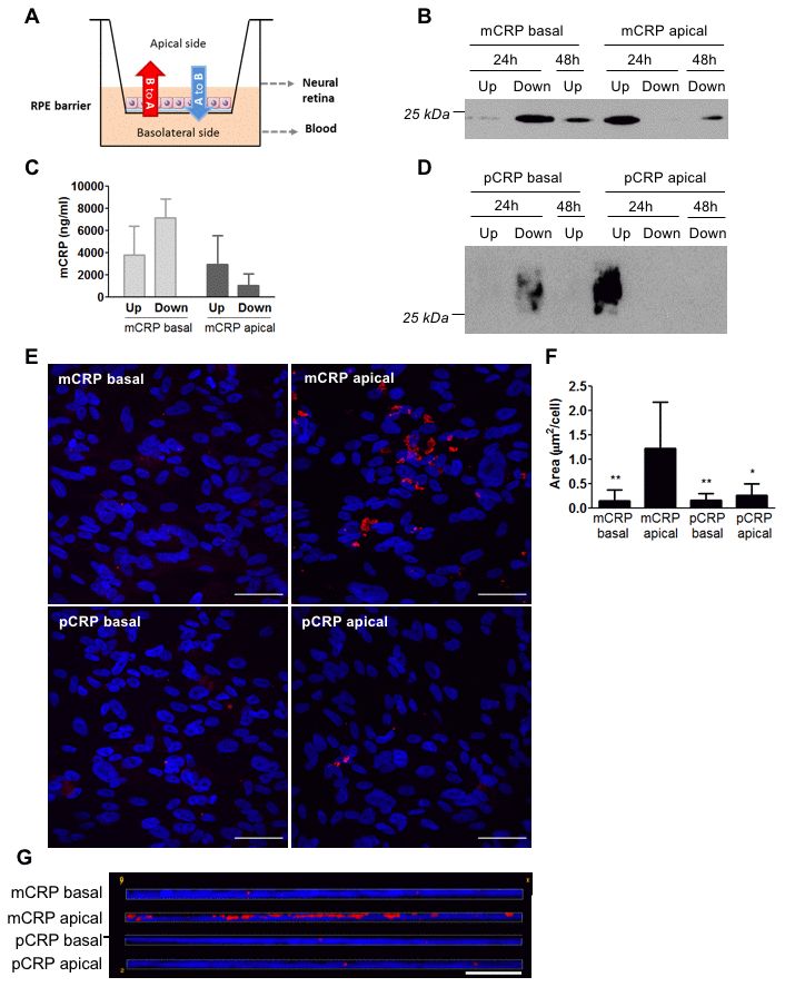
Figure 2.Diffusion of CRP isoforms across ARPE-19 cells. (A) Experimental setup. CRP (10 μg/ml) was added to either the apical or basolateral chamber of Transwell for 48h, mimicking neural retina and choriocapillaris, respectively. The presence of CRP in the opposite chamber where it was added was determined by Western blot and ELISA, and CRP bound to the cell surface was determined by immunofluorescence. (B) Western blot of mCRP present in apical (Up) and basolateral (Down) supernatants after 24 and 48 hours of treatment (N=4). (C) ELISA of mCRP (ng/ml) from apical (Up) and basolateral (Down) supernatants 48 hours after treatment. Values are expressed as mean ± SD (N=3). (D) Western blot of pCRP present in apical (Up) and basolateral (Down) supernatants after 24 and 48 hours of treatment (N=4). (E) Immunofluorescence of CRP (red) stained with monoclonal antibodies against mCRP (3H12) or pCRP (1C6). Nuclei stained with DAPI. Scale bar = 50 μm (N=6). (F) Quantification of CRP binding measured as stained area divided by the number of cells per image (μm2/cell). Results are expressed as mean area (μm2/cell) ± SD. Statistical analysis was performed by One-Way ANOVA and Tukey’s posthoc. * P<0.05, ** P<0.01 vs. mCRP apical. (G) Reconstruction of x-z sections with a 0.3 μm z axis step of immunofluorescence images. Images shown are representative of six independent experiments.