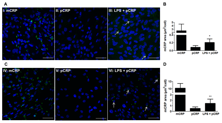
Figure 4.LPS-induced inflammation promotes CRP dissociation in RPE cells. RPE cells were treated with 100 μg/mL LPS for 24h before adding pCRP. After 24h, RPE cells were treated with pCRP for 48h and the presence of mCRP on the surface of RPE cells was measured by immunofluorescence. mCRP immunostaining of ARPE-19 (A) and primary porcine RPE (B) cells treated with 10 μg/ml mCRP for 48h (I, IV), 25 μg/ml pCRP for 48h (II, V), or 100 μg/ml LPS 24h before treatment with 25 μg/ml pCRP for 48h (III, VI). Arrows point mCRP dissociated from pCRP on RPE surface. Nuclei stained with DAPI. Scale bar = 50 μm. Images shown are representative of three independent experiments. (C, D) Quantification of CRP dissociation measured as stained area with the monoclonal antibody 3H12 against mCRP (green) divided by the number of cells per image (μm2/cell). Results are expressed as mean area (μm2/cell) ± SD (N=3). Statistical analysis was performed by student t-test. *P<0.05 vs. pCRP.