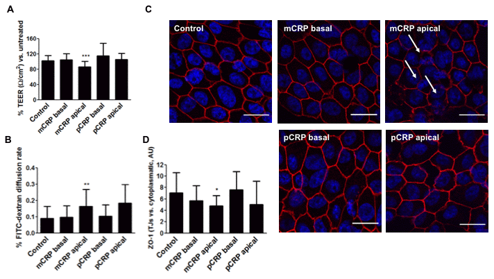
Figure 6.mCRP induces barrier disruption in primary porcine RPE cells in a polarized manner. Primary porcine RPE cells were treated with CRP isoforms for 48h either from the apical side or the basolateral chamber and TEER (A) and paracellular permeability as determined by FITC-dextran diffusion rate (B) was determined. (C) Cells were then fixed and immunostained with anti ZO-1 (red) and DAPI (blue). Images shown are representative of four independent experiments. Arrows show disruption of ZO-1. Scale bar = 20 μm. (D) Quantification of ZO-1 at the TJs expressed as relative (intercellular/cytoplasmic) ZO-1 distribution. Values are expressed as mean ± SD and statistical analysis was performed by one-way ANOVA and Dunnett´s posthoc analysis (N=6). * P<0.05, ** P<0.01, *** P<0.0001 vs. control.