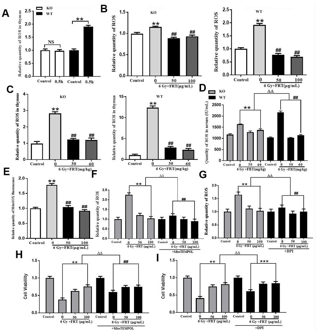
Figure 3.Effect of FRT on scavenging intracellular ROS due to PARP-1. (A) Representative flow cytometry histogram illustrating the radiation-induced change in ROS levels as detected by the DCFH-DA probe in thymus cells 0.5 h after radiation. (NS, not significant; ** P <0.01). Data was expressed as mean ±SD, n=5. (B) ROS levels in thymus cells measured by DCFH-DA probe. The thymus cells were irradiated at a dose of 6 Gy 2 hours after pretreatment with FRT (50 and 100 μg/mL). Cells were collected 0.5 h after irradiation. (** P<0.01 compared with normal group; ## P<0.01 compared with radiation group). Data was expressed as mean ±SD, n=5. (C) ROS levels in mice measured using DCFH-DA probe. Mice were pretreated with FRT (30 and 60 mg/kg) 4 days prior to 6 Gy irradiation, then sacrificed and thymus harvested 4 days after radiation. (**P <0.01 compared with normal group; ## P<0.01 compared with radiation group). Data was expressed as mean ±SD, n=5. (D) ROS levels in serum were quantified by ELISA 4 days after irradiation. (** P<0.01; ## P<0.01; ΔΔ P<0.01). Data was expressed as mean ±SD, n=5. (E) Representative luciferase labeling instrument displaying MitoSOX fluorescence in thymus cells under different conditions. Mitochondrial ROS as measured with the MitoSOX probe (**P<0.01 compared with normal group; ## P<0.01 compared with radiation group). Data was expressed as mean ±SD, n=5. (F) Thymus cells were treated with FRT 2 h before irradiation. The cells were incubated with MitoTEMPOL (50 μM) inhibitor for 1 h before irradiation then collected by centrifugation 0.5 h after irradiation. Flow cytometry was used to measure intracellular ROS levels. (** P<0.01; ## P<0.01; ΔΔ P<0.01). Data was expressed as mean ±SD, n=5. (G) Thymus cells were pretreated with FRT for 2 h before irradiation. The cells were incubated with DPI (10 μM) for 1 h prior to irradiation and 0.5 h after irradiation. The effect of DPI on ROS levels in thymus cells after irradiation was measured by flow cytometry. (** P<0.01; ## P<0.01; ΔΔ P<0.01). Data was expressed as mean ±SD, n=5. (H) The survival fraction of thymus cells treated with or without ROS inhibitor in mitochondria (MitoTEMPOL, 50 μM). Cell viability was measured 6 h after radiation by CCK-8 assay. MitoTEMPOL reduced the sensitivity of thymus cells to radiation and enhanced the viability of thymus cells (** P<0.01; ## P<0.01; ΔΔ P<0.01). Data was expressed as mean ±SD, n=5. (I) Proportion of surviving thymus cells treated with or without inhibitor of NAPDH oxidase (DPI, 10 μM). Cell viability was measured 6 h after radiation by CCK-8 assay. DPI reduced the sensitivity of thymus cells to radiation and enhanced their viability (** P<0.01; ## P<0.01; ΔΔ P<0.01). Data was expressed as mean ±SD, n=5.