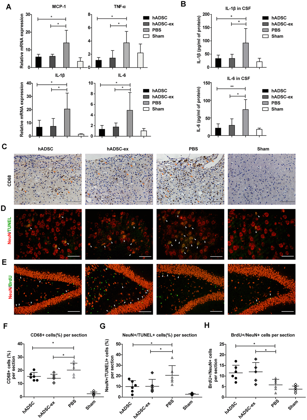
Figure 2.hADSC-ex suppressed neuroinflammation, reduced neuronal loss in the lesion boundary area and promoted hippocampal neurogenesis. (A) qRT-PCR analysis of proinflammatory factors in damaged brain tissues on day 5. (B) ELISA analysis of IL-1β and IL-6 levels in CSF on day 5. (C) CD68 immunohistochemical staining for activated microglia/macrophages (indicated by yellow arrows) in the lesion boundary zone on day 35. Scale bar = 100 μm. (D) NeuN immunofluorescence staining for mature neurons and TUNEL staining for apoptotic cells in the lesion boundary zone on day 14; double-staining with TUNEL (green)/NeuN (red) for apoptotic neurons is indicated by white arrows. Scale bar = 50 μm. (E) NeuN immunofluorescence staining for mature neurons and BrdU staining for cell proliferation in the hippocampal dentate gyrus on day 35; double-staining with BrdU (green)/NeuN (red) for newly generated mature neurons is indicated by white arrows. Scale bar = 100 μm. (F– H) Scatter plots of data from C, D and E. Data represent the mean ± SD, n = 6 rats per group. ns. p > 0.05, * p < 0.05, determined by one-way ANOVA vs. PBS control group.