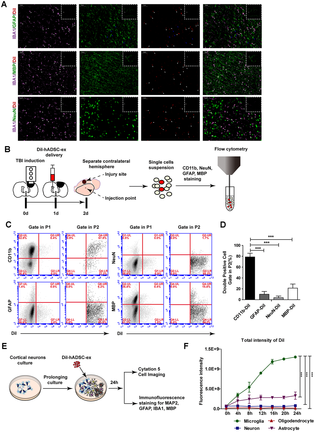
Figure 4.hADSC-ex were mainly taken up by microglia/macrophages in vitro and in vivo. (A) Representative images of IBA1/GFAP/DiI, IBA1/MBP/DiI and IBA1/NeuN/DiI immunostaining in the lesion boundary zone in rat brain coronal sections (bregma, −1.5 mm); n = 3, scale bar = 50 μm. The overlapping signals are marked with blue arrows (GFAP/DiI) and white arrows (IBA1/DiI). The white dotted boxes denoted the slices overview and the solid line rectangles indicated the snapshot location. (B) Schematic representation of the experimental procedures to detect hADSC-ex cellular uptake by dissociated primary neural cells using FACS. (C) Representative dot plots from FACS showing double-positive cell (CD11b/DiI, GFAP/DiI, NeuN/DiI, MBP/DiI) gating in P1 and P2. The gating strategy is shown in Supplementary Figure 3A. (D) Bar graphs quantifying the data from (C). Data are presented as the mean ± SD, n = 3 independent experiments, *** p < 0.001, determined by one-way ANOVA vs. CD11b/DiI. (E) Schematic representation of the use of mixed neural cell cultures to identify the cellular uptake of hADSC-ex in vitro. (F) Line graph showing the change in the total fluorescence intensity of DiI over time in every neural cell type in the mixed neural cell culture. Data represent the mean ± SD, n = 3 independent experiments, *** p < 0.001, determined by repeated-measures two-way ANOVA vs. microglia.