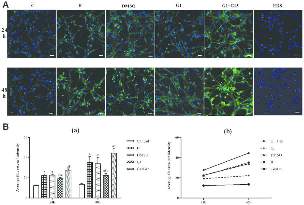
Figure 4.GPER activation reduces autophagy in hyperoxia-treated primary retinal microglia. (A) Representative confocal fluorescence images (400×, Bar=25μm) show Cyto-ID labeled autophagic vesicles (green) in the (a) control (C), (b) hyperoxia (H), (c) hyperoxia+DMSO (DMSO), (d) hyperoxia+G-1 (G-1), and (e) hyperoxia+G-1+G-15 (G-1+G-15) groups of primary retinal microglia at 24 and 48 h. The negative control cells were treated with PBS instead of Cyto-ID. (B) Histogram plots show average fluorescence staining intensity (Cyto-ID; green) at 24 and 48 h in the (a) control (C), (b) hyperoxia (H), (c) hyperoxia+DMSO (DMSO), (d) hyperoxia+G-1 (G-1), and (e) hyperoxia+G-1+G-15 (G-1+G-15) groups at 24 and 48 h. P<0.05 denotes statistical significance. The experiments were repeated thrice and means±S.D are shown.