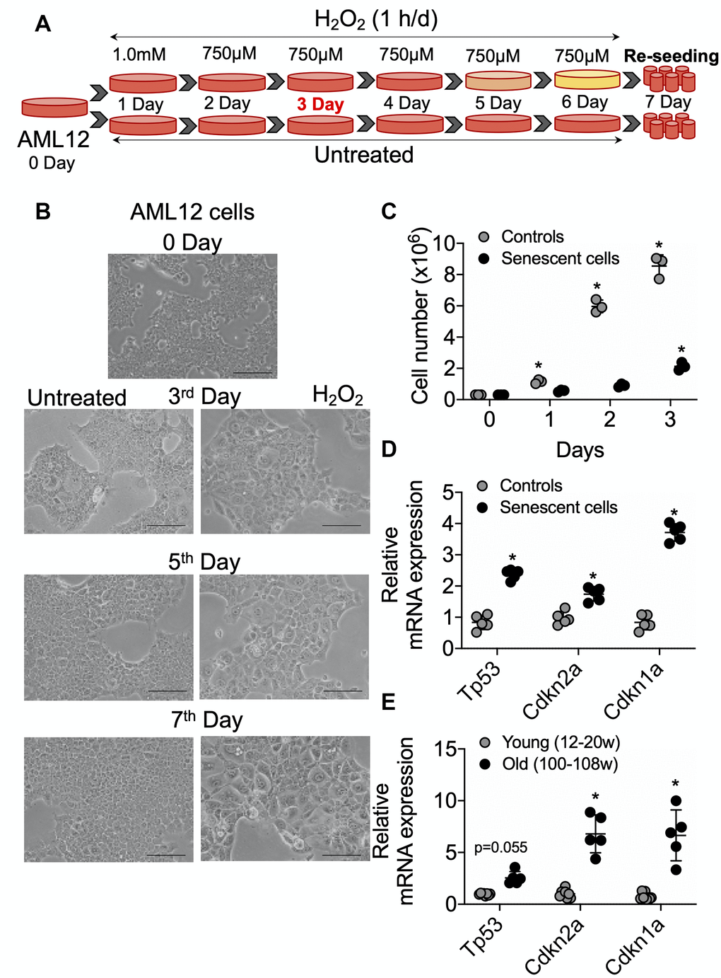
Figure 1.Senescence induction in mice normal hepatic cells AML12. (A) Schematic representation of experimental strategy for the senescence induction. 4x10⁶ Cells were seeded at day 0 in the T175 cell culture flask. Next day (day 1) cells were treated with 1.0 mM H2O2 followed by 750 μM for the subsequent 5 days. Cells can be sub-cultured at 1:3 ratio on day 3 if required. At day 7, cells were re-seeded for experiments as required. (B) Visualization of senescence induction in mice AML12 cells during H2O2 treatments from day 3, day 5, and day 7 before re-seeding. The images are taken randomly at 10x magnification. Scale bars as 100 μm. (C) 0.3x10⁶ control and senescent AML12 cells were seeded in each well of the 6-well plate. Cells were trypsinized at indicated days and counted by an automated cell counter. (D and E) RT-qPCR analysis of senescence genes in AML12 cells (D) and liver tissues from young and old mice (E). * Statistical differences were calculated significant as p<0.05.