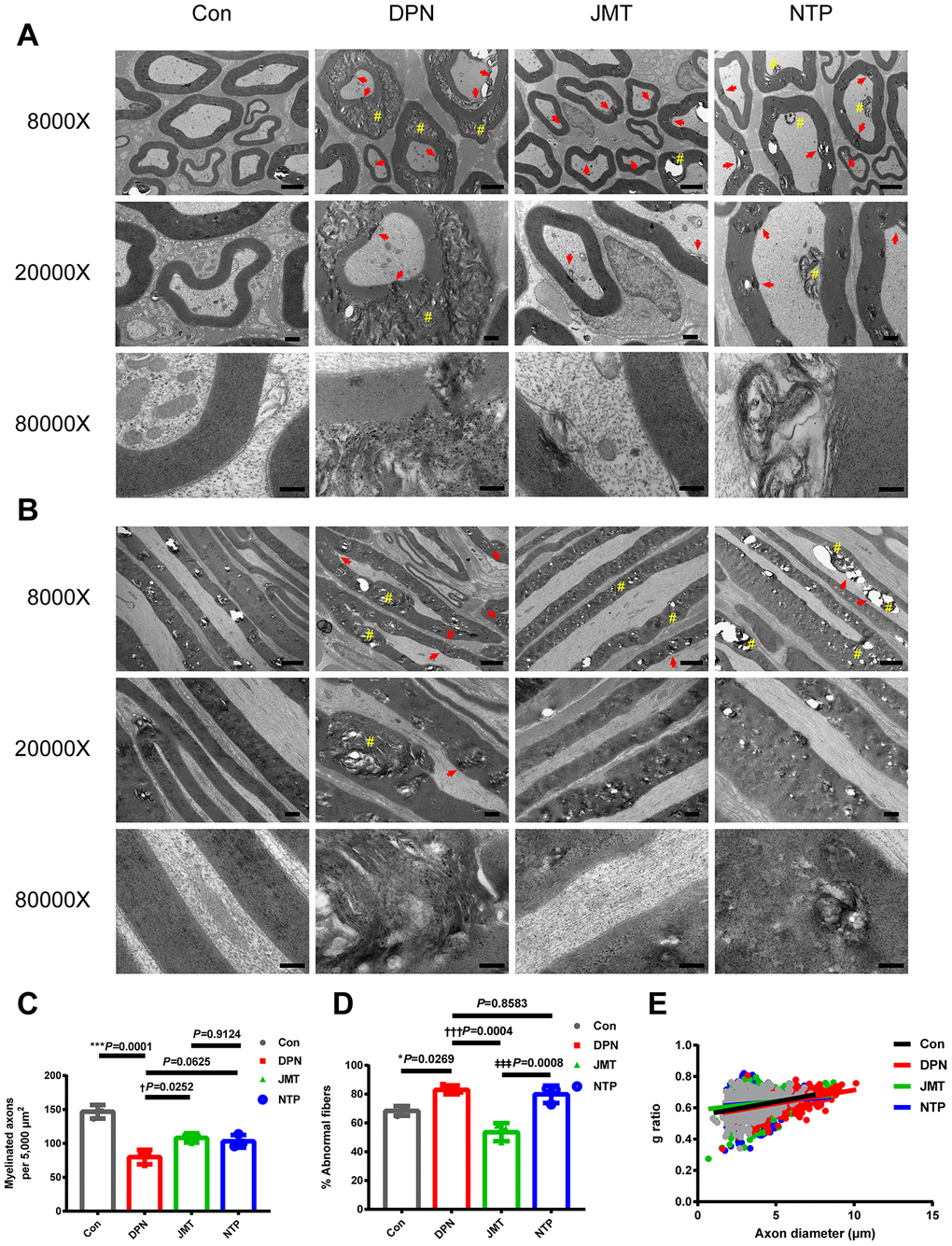
Figure 2.Ultrastructure and morphometry of sciatic nerves under transmission electron microscopy. Representative images of (A) cross-sections and (B) longitudinal sections of sciatic nerves from different groups at magnifications of 8000×, 20000×, and 80000×; scale bars, 2 μm, 500 nm, and 200 nm, respectively. Morphometric analyses of sciatic nerve showing (C) the number of myelinated axons per 5,000 μm2 and (D) the percentage of abnormal myelin fibers in different groups. Means ± SD; n = 3/group. (E) Quantification of g ratios for axons in different groups. n=632 axons from 3 normal control rats, n=525 axons from 3 distilled water-treated DPN rats, n=585 axons from JMT-treated DPN rats, n=544 axons from 3 NTP-treated DPN rats. ***p < 0.001, *p < 0.05, vs. normal control group; †††p < 0.001, †p < 0.05 vs. distilled water-treated DPN group; ‡‡‡p < 0.001 vs. JMT-treated DPN group. One-way ANOVA followed by Tukey’s multiple comparison test or Kruskal-Wallis test followed by Dunn’s multiple comparisons test. Pound signs (#) indicate onion-bulb and bubble form protrusions. Red arrows indicate lamellar separation between the axon and myelin sheath and demyelination.