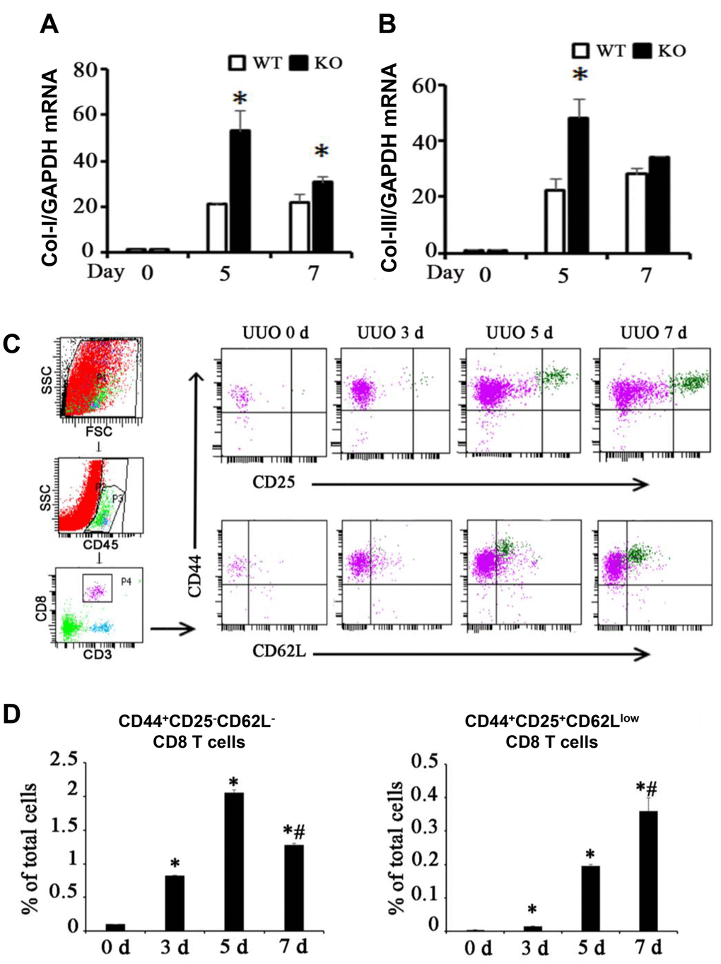
Figure 1.Two different subsets of infiltrating CD8 T cells were related to renal fibrosis in UUO model mice. (A, B) Total mRNA was obtained from the UUO kidneys of WT mice, CD8 KO mice, or CD8 KO mice transplanted with CD8 T cells at days 0, 5, and 7. The results showed the expression levels of Col-I and Col-III in CD8 KO mice compared with those in WT mice. (*p < 0.05 vs. WT UUO). (C) Representative examples of FACS analysis at each point. CD8+ T cells in obstructed kidneys were identified on the basis of CD45, CD3, CD44, CD25, and CD62L expression by using flow cytometry. (D) CD44+CD25−CD62L− and CD44+CD25+CD62Llow CD8+ T cells were counted and analyzed at days 0, 3, 5, and 7 after UUO (*p < 0.05 vs. 0d, #p < 0.05 vs. 5d).