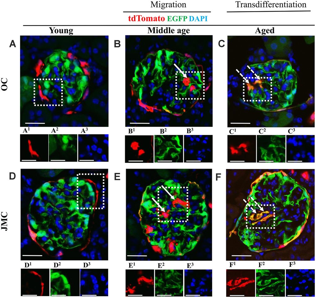
Figure 2.Migration and differentiation of tdTomato+PECs to a podocyte phenotype in aged kidneys. (A–F) Representative confocal images of tdTomato-labeled PECs (red), EGFP+ labeled podocytes (green), and DAPI stained nuclei (blue) in young, middle age and aged mice in OC (A–C) and JMC (D–F). Individual red (labeled with superscript-1), green (superscript-2) and far-red (superscript-3) fluorescent channels of the glomerulus, outlined by the white dashed box. (A) Young mice (OC) showed that tdTomato+PECs are detected along Bowman’s capsule (A1) and EGFP+ cells (A2) localized in typical podocyte distribution. Nuclei were labeled with DAPI (A3). (B) Middle age mice (OC) showed that tdTomato+PECs (marked with solid arrow) were detected in the glomerular tuft (B1) with accompanied decrease in the EGFP signal (B2) and the nuclear marker DAPI (B3). (C) Aged mice (OC) showed that tdTomato+PECs (marked with dashed arrow) (C1) differentiated to a podocyte fate and co-expresses EGFP (green) (C2), overlap creates a yellow. (D) Young mice (JMC) showed that tdTomato+PECs are detected along Bowman’s capsule (D1) and EGFP+ podocytes are observed in the glomerular tuft (D2). (E) Middle age mice (JMC) showed that tdTomato+PECs (marked with solid arrow) are observed in the glomerular tuft (E1) with no overlap with EGFP (E2). (F) Aged mice (JMC) showed that tdTomato+PECs (marked with dashed arrow) (F1) overlap with EGFP (F2) and create a yellow color in the glomerular tuft. Scale bars represent 25μm or 5μm (insets).