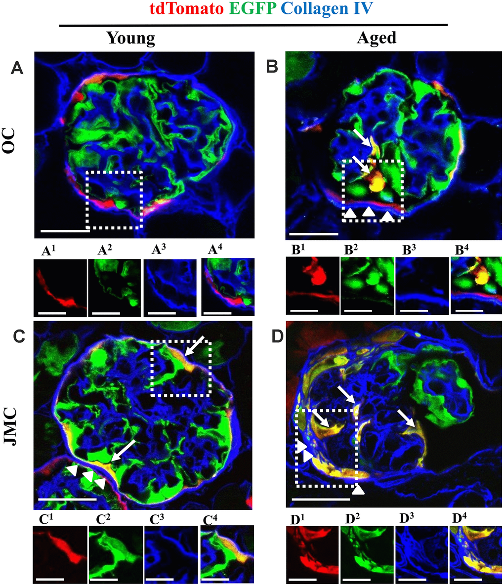
Figure 4.Collagen IV staining increases along Bowman's capsule with accompanied migration of tdTomato+PECs in aged mice. (A–D) Representative confocal images of tdTomato+ (red), EGFP+ (green) and Collagen IV staining (blue) in young and aged mice. The inserts show separate channels of the outlined glomeruli, with superscripts: 1=tdTomato, 2=EGFP, 3=Collagen IV and 4=merged. (A) Young mice (OC) showed that tdTomato+PECs are limited to Bowman’s capsule (A1) and EGFP+ podocytes to the glomerular tuft (A2). Collagen IV staining (blue) (A3) was used to delineate the kidney architecture. (B) Aged mice (OC) showed that differentiated yellow cells (tdTomato+PECs+ EGFP+) (marked with white arrows) were detected in the glomerular tuft (B1, B2) which was accompanied by an increase in Collagen IV staining along BC (white arrow heads) (B3). (C) Young mice (JMC) showed that differentiated yellow cells (tdTomato+PECs+ EGFP+) were detected in some glomeruli along Bowman’s capsule (C1, C2) which was accompanied by an increase in Collagen IV staining along BC (white arrow heads) (C3). (D) Aged mice (JMC) showed that differentiated yellow cells (tdTomato+PECs+ EGFP+) have migrated to the glomerular tuft (marked with white arrows) and co-express tdTomato (D1), EGFP (D2), which was accompanied by an increase in Collagen IV staining along BC (white arrow heads) (D3). Scale bars represent 25μm or 5μm (insets).