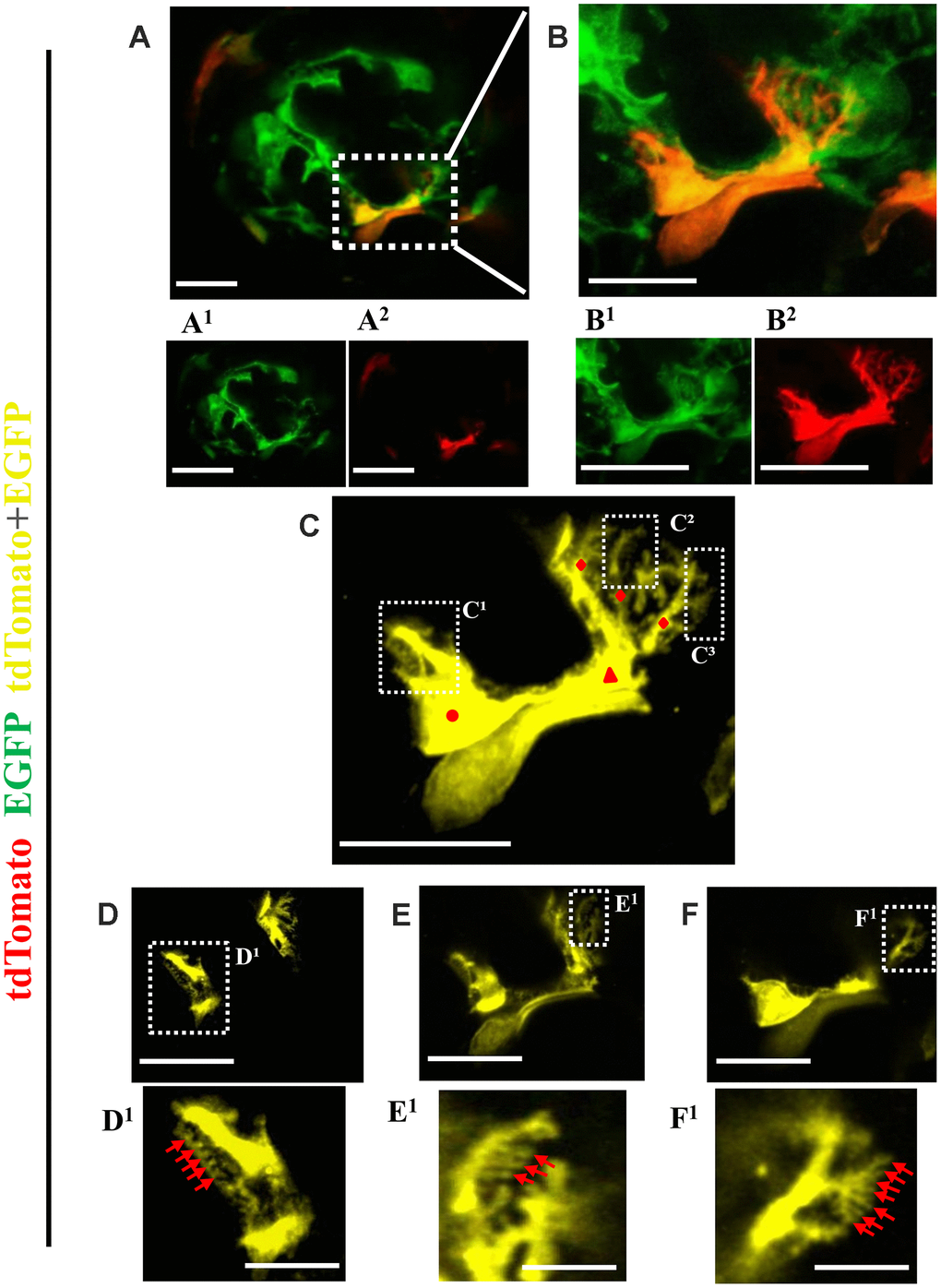
Figure 7.Ultrastructure of newly generated podocytes from PEC origin (tdTomato+EGFP+) in the glomerular tuft of aged mice. (A) Representative confocal image of expansion microscopy of a glomerulus in an aged mouse with a newly generated podocyte from PEC origin (marked with dashed box). Superscripts show separate channels: tdTomato+ (red) (A1) and EGFP+ (A2). (B) Higher magnification of image shown in A of newly generated podocyte, with accompanied single color images of tdTomato (B1) and EGFP (B2). (C) The newly generated podocyte has classic podocyte architecture: a cell body (labeled with red circle), primary process (red triangle), several secondary processes (red rhombuses), branching to minor and tertiary foot processes (C1–C3 labeled with dashed boxes). (D–F) Higher magnification of images shown in dashed boxes C1–C3 with different z-stack focal planes show various tertiary foot processes (labeled with red arrows). Scale bars represent 25μm or 5 μm (inset) and are in pre-expansion dimensions.