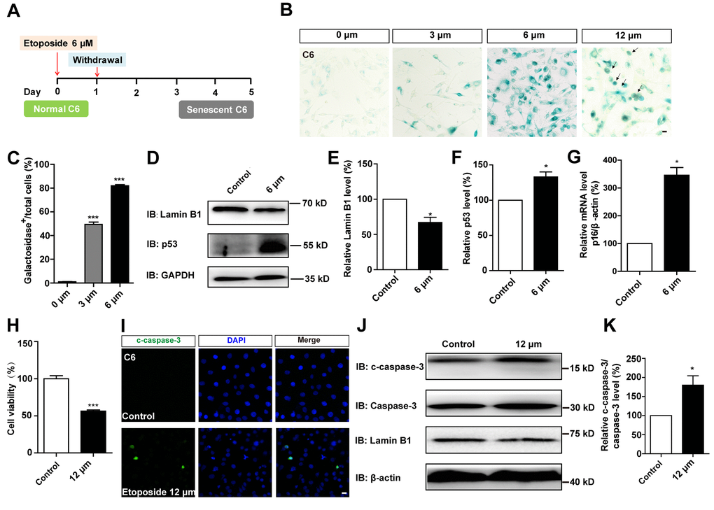
Figure 5.D-gal more effectively induced senescence of GBM cells than etoposide. (A) A schematic diagram showing etoposide-induced C6 cell senescence. (B) Representative images showing β-galactosidase staining in C6 cells treated with etoposide at 0 μM, 3 μM, 6 μM, 12 μM for 1 d, and recovered for 4 d. The black arrows indicated apparently dead cells. (C) Quantification of the percentage of β-galactosidase+ C6 cells over total cells as shown in (B) (n=15). (D) Western blot detected the expression of Lamin B1 and p53 in control cells and C6 cells treated with etoposide at 6 μM for 1 d, and recovered for 4 d. (E, F) Quantification of Lamin B1 and p53 expression as shown in (D) (n=5). (G) qPCR analysis for p16 mRNA level in control cells and senescent C6 cells (treated with etoposide at 6 μM for 1 d, and recovered for 4 d) (n=3). (H) The effects of etoposide (treated with 12 μM etoposide for 1 d, and recovered for 4 d) on the viability of C6 cells as detected by CCK8 assay (n=3). (I) Immunostaining of c-caspase-3 in control cells and senescent C6 cells (treated with 12 μM etoposide for 1 d, and recovered for 4 d). (J) Western blot detected the expression of c-caspase-3, caspase-3, and Lamin B1 in control cells and senescent C6 cells (treated with etoposide at 12 μM for 1 d, and recovered for 4 d). (K) Quantification of c-caspase-3/caspase-3 level as shown in (J) (n=4). Scale bars, 20 μm. Data shown are mean ± s.e.m. *P < 0.05, ***P < 0.001.