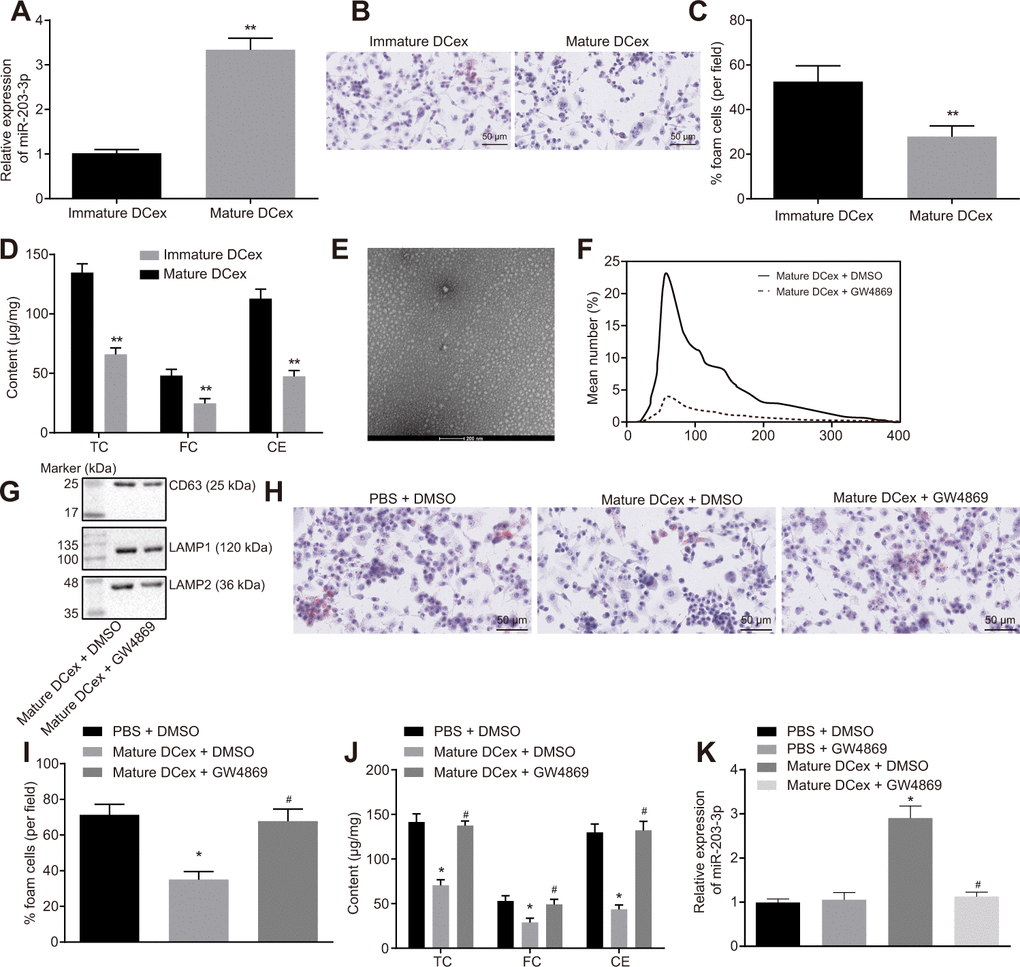
Figure 3.BMDM phenotypes of AS are repressed by DCex. BMDMs were initially treated with ox-LDL or without treatment. (A) miR-203-3p expression in immature and mature exosomes from DCs by RT-qPCR. (B, C) The proportion of foam cells in immature and mature exosomes from DCs by oil red O staining (200 ×). (D) TC, FC and CE levels in immature and mature exosomes from DCs determined by ELISA. (E) Morphology of exosomes after GW4869 treatment (scale Bar = 200 nm) detected by transmission electron microscopy. (F) Nanosight particle tracking analysis of the size and quantity of exosomes. (G) Western blot analysis of the expression of exosome marker proteins. (H–I) The proportion of foam cells in BMDMs after 24-h stimulation with ox-LDL measured by oil red O staining (200 ×). (J) Serum TC, FC and CE levels in BMDMs after 24-h stimulation with ox-LDL determined by ELISA. (K) miR-203-3p expression in BMDMs determined by RT-qPCR. *p < 0.05 and **p < 0.01 vs. the BMDMs without treatment; *p < 0.05 vs. PBS + DMSO; #p < 0.05 vs. DCex + DMSO. Statistical data were measurement data, and described as mean ± standard deviation. The paired t test was used for comparisons between two groups in panel (A–D). The one-way analysis of variance was adopted for comparisons among multiple groups in panel (I–K) followed by Tukey’s post hoc test. The experiment was repeated 3 times independently.