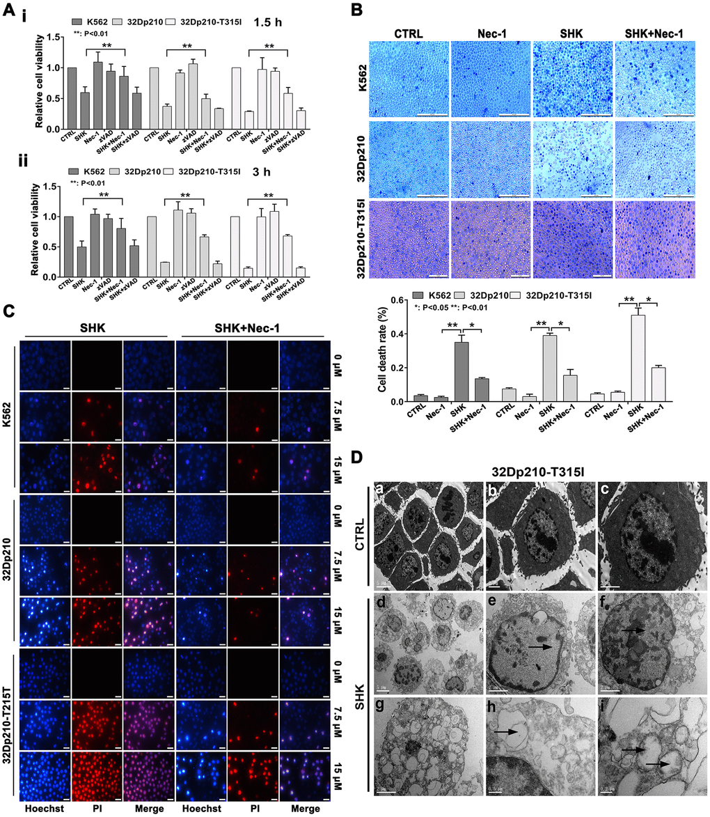
Figure 1.Shikonin induces necroptosis in CML cells. (A) Results of MTT proliferation assays in K562, 32Dp210, and 32Dp210-T315I CML cells treated with 10 μΜ shikonin for 1.5 h (i) or 3 h (ii) following pre-treatment (1 h) with Nec-1 (50 μΜ) or zVAD-fmk (35 μΜ). Data are presented as the mean ± SD of three independent experiments. (B) Trypan blue exclusion assay results. CML cells were pretreated with or without 50 μΜ Nec-1 for 1 h and subsequently exposed to 10 μΜ shikonin for 3 h. The cells were then stained with trypan blue and the percentage of dead cells was determined under light microscopy. (C) Hoechst 33342/PI double staining was performed in CML cells preincubated with or without 50 μΜ Nec-1 and then treated with 0, 7.5, or 15 μΜ shikonin for 3 h. The percentage of PI-permeable cells in each group was determined by fluorescence microscopy. Blue fluorescence indicates staining with Hoechst 33342 and red fluorescence indicates PI staining. A few cells exhibited apoptotic characteristics (chromatin condensation and nuclear fragmentation), as indicated by white arrowheads. Magnification, 200×. (D) Electron microscopic examination of 32Dp210-T315I cells treated with 20 μM shikonin for 3 h revealed typical necrotic changes, including disorganization and loss (empty bubble-like formations) of cytoplasmic structures and plasma membrane rupture (d, g, h); syncytial nuclei with chromatin dissolution and disappearance of nucleoli (e, f); and severe damage to mitochondria with disruption of internal structures (i). Scale bars: 5 μm (a, d), 2 μm (b, c, e, f, g), 0.5 μm (h), and 0.2 μm (i). Quantification data are presented as the mean ± SD of three independent experiments. Representative results from three samples are shown. *p < 0.05; **p < 0.01.