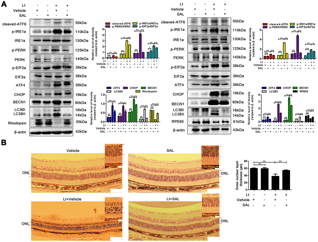
Figure 10.Suppressing ER stress inhibits prolonged autophagy and protects the retina against light injury. The mice were intraperitoneally injected with a dose of 1 mg/kg once a day for 7 days. On the third day of administration, the mice were exposed to continuous 7000 Lux visible light for 12 h. After light exposure, the mice were fed in the animal room with the normal light/dark cycle. On the fifth day of light exposure, the mice were sacrificed, and the eyeballs were enucleated. (A) The retinas were collected, and target proteins were determined with western blotting. β-actin was referenced as an internal control. Three independent experiments are conducted three weeks apart. The results are presented as the mean± SEM. n (per group) =3, NS: no significance, *P < 0.05, **P < 0.01. (B) The retinas were sectioned and stained with H&E and photographed under a microscope. Scale bar=100 μm; 20 μm. The thickness of the outer nuclear layer (ONL) was measured and quantitatively analyzed. The results are presented as the mean± SEM, n (per group) =6, NS: no significance, **P < 0.01.