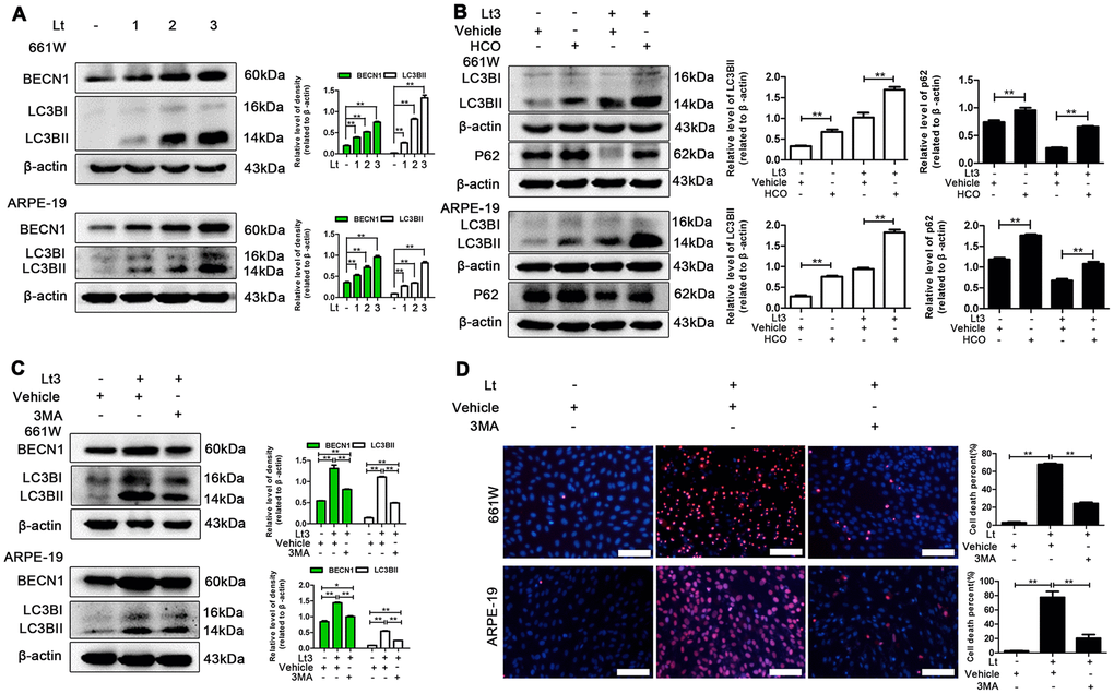
Figure 6.Inhibiting light-induced prolonged autophagy is protective. (A) 661W cells/ARPE-19 cells were cultured in a dark condition or exposed to 1500 Lux light for 1–3 days. The levels of BECN1 and LC3BII in the whole cell lysate was determined with western blotting, and β-actin was referenced as an internal control. (B) After 661W cells and ARPE-19 cells were treated with HCO (20 μM) or vehicle and cultured under light/dark conditions for 3 days, the level of LC3BII and P62 in the whole cell lysate were determined with western blotting, and β-actin was referenced as an internal control. (C) The cells were treated with 3MA (2.5 mM for 661W cells; 1 mM for ARPE-19) or vehicle and cultured under light/dark conditions for 3 days. The level of BECN1 and LC3BII in the whole cell lysate were determined with western blotting, and β-actin was referenced as an internal control. (D) 661W cells pretreated with 2.5 mM 3MA/vehicle were cultured under light/dark conditions for 3 days. ARPE-19 cells pretreated with 1 mM 3MA/vehicle were cultured under light/dark conditions for 6 days. The percentage of cell death was evaluated with PI/Hoechst staining. Scale bar=100 μm. Three independent experiments are conducted two weeks apart. The results are presented as the mean± SEM. n (per group) =3, *P < 0.05, **P < 0.01.