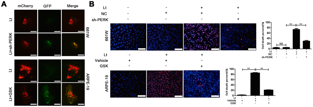
Figure 9.Inhibiting PERK blocks autophagic flow and protects the light-damaged cells. (A) 661W cells with stable PERK knockdown and ARPE-19 cells were infected with mCherry-GFP double labeled-LC3B mediated by adenovirus. At 48 h after infection, the ARPE-19 cells were treated with GSK (5 μM) or vehicle. The cells were cultured under 1500 Lux light condition for 3 days and photographed under fluorescence microscopy. Scale bar=20 μm. (B) 661W cells with PERK knockdown were cultured under light/dark conditions for 3 days, but ARPE-19 cells treated with GSK (5 μM) or vehicle were cultured under light/dark conditions for 6 days. The percentage of cell death was evaluated with PI/Hoechst staining. Scale bar=100 μm. Three independent experiments are conducted two weeks apart. The results are presented as the mean± SEM. n (per group) =3, NS: no significance, **P < 0.01.