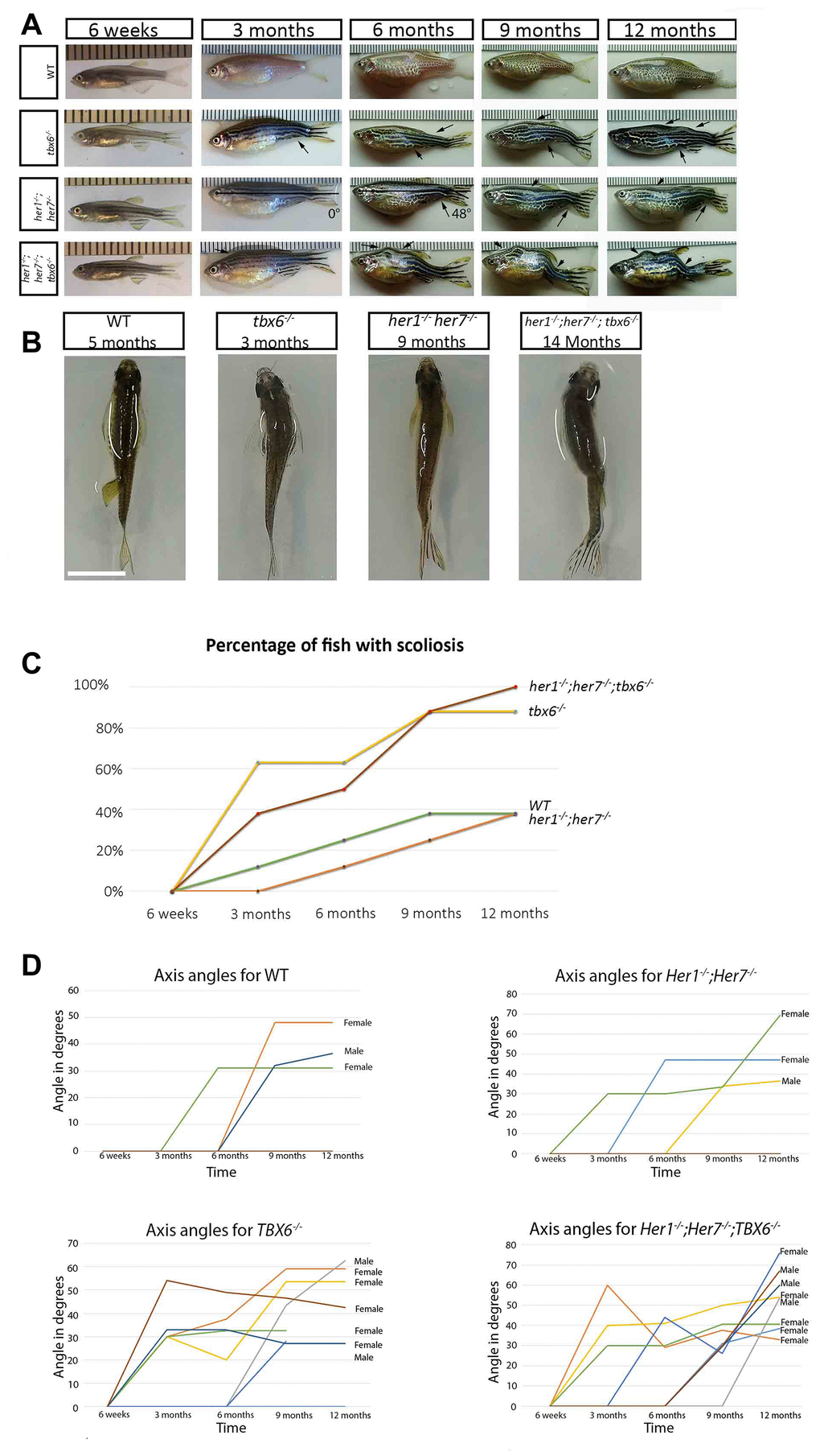
Figure 2.Clock segmentation mutants develop adult scoliosis. (A) Representative time lapse images of individuals from each genotype over the period from 6 weeks to 12 months, allowing to track the development of scoliosis (arrow). Mutant individuals have already mild signs of scoliosis at 3 months, while wild type exhibit the first indication of deviation from the body axis at 6 months. In the her1-/-; her7-/- individual at 3 and 6 months is an exemplary depiction of how scoliosis measurements were carried out. (B) Dorsal view of the different genotypes, showing an S body shape characteristic for scoliosis. (C) Graphical representation of the percentage of fish developing scoliosis over time, reaching 100% in the triple mutants and 83% in the tbx6-/- at the end point. In the wild type and in the her1-/-; her7-/- mutants only 38% presented scoliosis. (D) Measurements of axis angles in different individuals at different time points during virtual time-lapse. Only the individuals with an angle of deviation from the body axis are shown in the graph, none bended individuals have an angle of zero. Different line colours represent individual fish. Between 9 and 12 months, two wild type fish (1 with scoliosis and 1 without scoliosis), one her1-/-; her7-/- (with scoliosis) and two tbx6-/- (both with scoliosis) had to be sacrificed. After the individual was removed, it was still counted as bended or normal in the 12-month quantification. Note: the angle can decrease or increase depending on how the angle of deviation from the body axis develops over time in the individual. The ruler in section A serves as a scale bar, the space between two successive lines marks one millimetre. The scale bar in section B represents 1 cm.