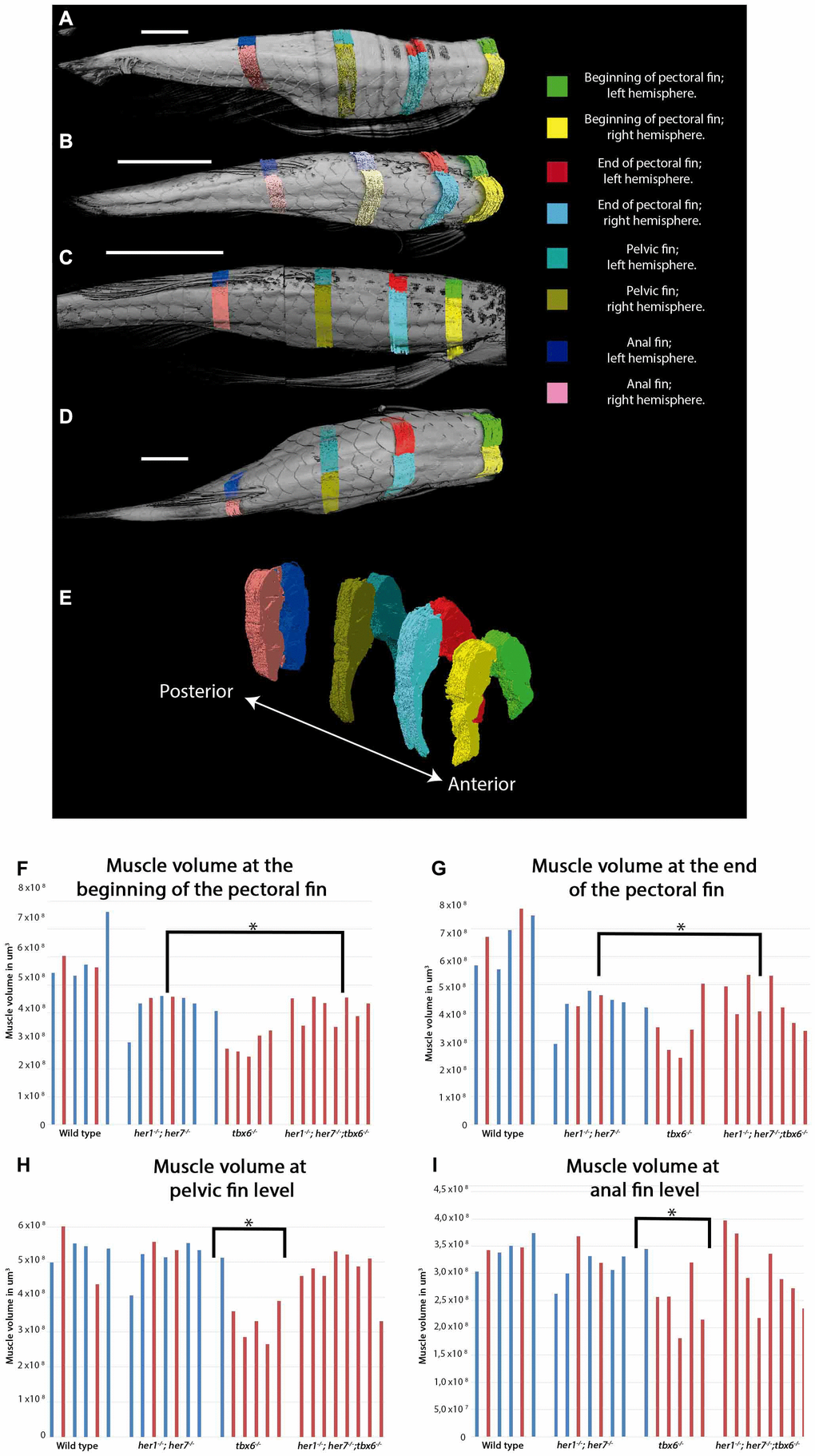
Figure 3.Muscle volume is affected in mutants at adult stages. (A–D) Representative micro CT images showing the dorsal view of fish used for the segmentation of muscles into the left and right side at the four key areas (indicated by the colored regions) in the 4 different groups: (A) wild type, (B) tbx6-/- (C) her1-/-;her7-/-, and (D) her1-/-;her7-/-;tbx6-/-. (E) Lateral view of reconstructed surface generation of the individual muscle from the WT. (F–I) graphical results of the volume measurements for every area. (F) Muscle volume at the beginning of the pectoral fin. (G) Muscle volume at the end of the pectoral fin. (H) Muscle volume at pelvic fin level. (I) Muscle volume at anal fin level. An asterisk denotes a statistically significant difference between wild type and mutant groups (two tailed significant test P=0,05). Individuals that presented scoliosis during the course of the experiment (represented with red bars) tend to have the same muscle volume as their phenotypically normal siblings (represented with blue bars). The three mutants analyzed have less muscle volume at the first two anterior positions. The muscle of 27 out of 32 individuals could be analyzed to 12 months, because two wild type fish (1 with scoliosis and 1 without scoliosis), one her1-/-; her7-/- individual (with scoliosis) and two tbx6-/- individuals (both with scoliosis) had to be euthanized between 9 months and one year. After the individual was removed, it was not stained for muscle analysis and therefore will not appear in the graph.