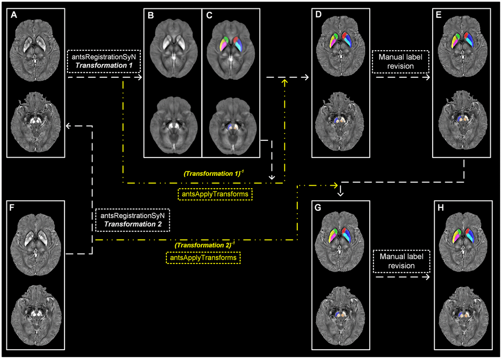
Figure 3.Semi-automatic extraction of regional tissue susceptibility. (A) Native QSM image at baseline; (B, C) QSM template ant the subcortical label in its space; (D) Warped subcortical label in the native QSM image at baseline; (E) Final label in the data analysis through manual revision; (F) Native QSM image at follow-up; (G) Warped subcortical label in the native QSM image at follow-up; (H) Final label in the longitudinal data analysis through manual revision. ANTs-SyN algorithms are used to complete the image coregistrations. QSM = Quantitative susceptibility mapping.