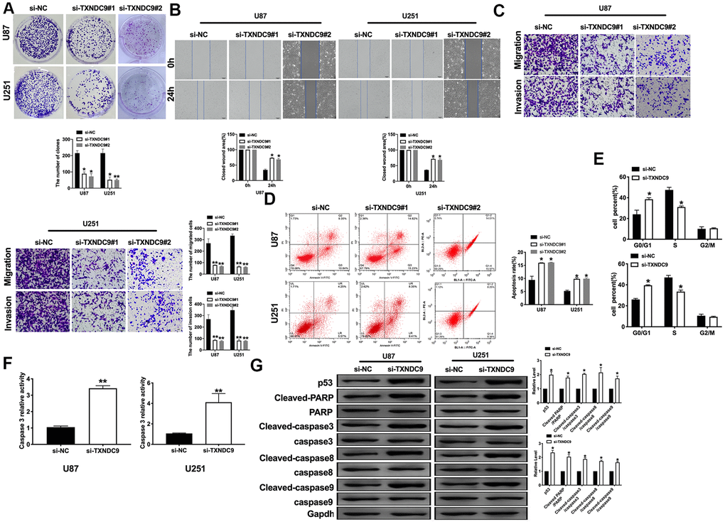
Figure 2.Knockdown of TXNDC9 prevented proliferation and induced apoptosis of U87 cells. (A) The colony formation assay. (B, C) Wound healing assay and transwell were performed for detecting the effect of TXNDC9 on migration and invasion. (D) The apoptosis rate of U87 and U251 cells were measured by flow cytometry. The histogram at the right is a statistical graph. n=4, *P<0.05. (E) Flow cytometry was performed to determine the cell cycle in U87 and U251 cells after transfection si-TXNDC9/si-NC. n= 4, *P<0.05. (F) The caspase3 activity of U87 and U251 cells was evaluated by the caspase3 activity kit. n= 6, **P<0.01. (G) The protein level of p53, Cleaved-caspase3, Cleaved-caspase8, and Cleaved-caspase9 were detected by western blot, Gapdh was indicated as a loading control. n= 6, *P<0.05.