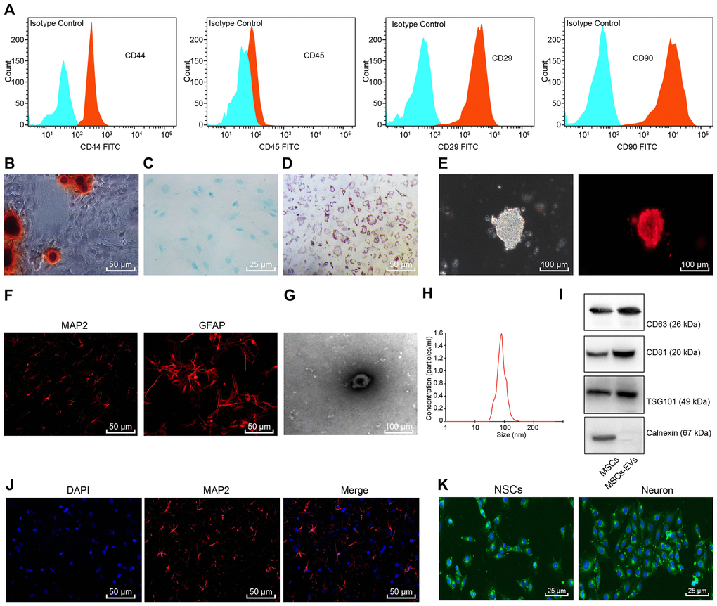
Figure 1.Identification of BMSCs, NSCs, and BMSCs-derived EVs. (A) Immunophenotypic analysis for BMSCs using flow cytometry. Blue represents Isotype control and red represents CD44+/CD45+/CD29+/CD90+ cells. (B) Alizarin red staining of BMSCs (× 200). (C) Alsine blue staining of BMSCs (× 400). (D) Oil red O staining of BMSCs (× 200). (E) Morphology of rat NSCs and Nestin-positive cells detected using immunofluorescence staining (scale bar = 100 μm). (F) The expression of MAP-2 and GFAP in the differentiated NSCs detected using immunofluorescence staining (× 200). (G) Morphology of EVs observed under TEM (scale bar = 100 nm). (H) Particle size distribution of BMSCs-derived EVs measured by NTA. (I) The contents of the surface markers CD81, CD63 and TSG101 in BMSCs and BMSCs-derived EVs measured using Western blot analysis. (J) Neuronal marker MAP-2 detected by immunofluorescence staining (× 200). (K) The uptake of EVs observed under the fluorescence microscope (× 400); green corresponded with the PKH-67-labelled EVs, and blue corresponded with the DAPI-stained BMSCs.