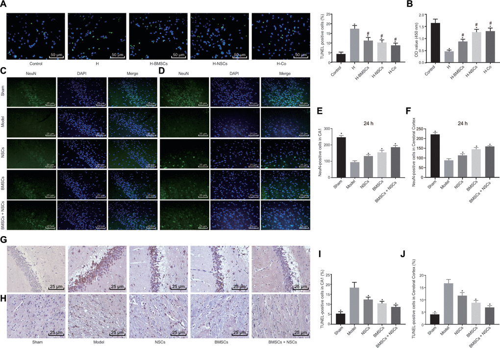
Figure 2.Combined treatment of BMSCs and NSCs promotes survival of neuronal cells in vivo and in vitro. Neuronal cells injured by hypoxia were untreated (H) or co-cultured with BMSCs (H-BMSCs), NSCs (H-NSCs) or BMSCs + NSCs (H-Co) in Panels (A and B). The sham-operated rats were not treated with any cells (sham) while CA rats were not treated (Model) or injected with BMSCs, NSCs or BMSCs + NSCs in Panels (C–J). (A) Apoptosis of neuronal cells assessed by TUNEL staining (× 200). (B) Cell viability assessed by CCK-8 assay. (C) Representative images of the NeuN-positive cells in the hippocampal CA1 region visualized using immunofluorescence staining (× 400). (D) Representative images of NeuN-positive cells in the cerebral cortex visualized using immunofluorescence staining (× 400). (E) The number of NeuN-positive cells in the hippocampal CA1 region. (F) The number of NeuN-positive cells in the cerebral cortex. (G) Apoptosis of neuronal cells in the hippocampal CA1 region assessed by TUNEL staining (× 400). (H) Apoptosis of neuronal cells in the cerebral cortex assessed by TUNEL staining (× 400). (I) Comparison of apoptotic rate in the hippocampal CA1 region. (J) Comparison of apoptotic rate in the cerebral cortex. * p < 0.05 vs. the Control (neuronal cells without any treatment) or Model (rats with CA without any treatment) group, # p < 0.05 vs. the H group (hypoxia-induced injured neuronal cells without any treatment). Data were expressed as mean ± standard deviation, and comparison among multiple groups were analyzed by one-way ANOVA followed by Tukey's post hoc test. n = 10 in animal experiments. The cell experiments were conducted 3 times independently.