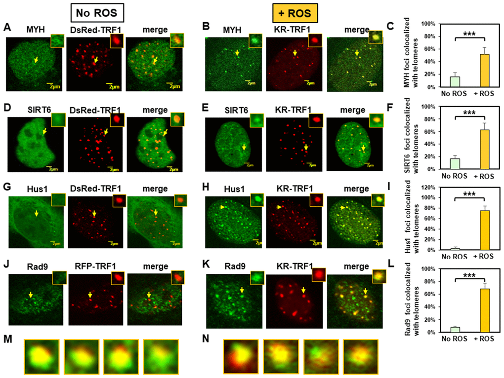
Figure 1.Expressed MYH, SIRT6, Hus1, and Rad9 are recruited to oxidatively damaged telomeric sites in mouse embryonic fibroblast (MEF) cells. (A), (D), (G), and (J), Distribution of GFP-hMYH, GFP-hSIRT6, GFP-hHus1, and FLAG-Rad9, respectively, in undamaged MEF cells containing DsRed-TRF1 or RFP-TRF1. Some GFP-hMYH and GFP-hSIRT6 granules are localized to DsRed-TRF1 at telomeres as shown in enlarged merged images. (B), (E), (H), and (K), GFP-hMYH, GFP-hSIRT6, GFP-hHus1, and FLAG-Rad9, respectively, form foci at KR-TRF1 damaged telomeric sites after light activation. Images were captured 30 min after light activation with an Olympus FV1000 confocal microscopy system. (C), (F), (I), and (L), Quantitative analyses of 20 cells in each undamaged and KR-induced damaged group. About 15% of granulated GFP-MYH and GFP-SIRT6 spots were localized to DsRed-TRF1 at telomeres (C, F). Over 50% of GFP-MYH, GFP-SIRT6, or GFP-Hus1 foci are colocalized with KR-TRF1 after KR activation. Error bars indicate SD; n ≥ 20. The P-value is calculated by Student’s t-test using Stat Plus software; P < 0.01 is shown as ***. (M), Enlarged merged images from (H) showing segregation of green GFP-hHus1 and red KR-TRF1 foci. (N), Enlarged merged images from (K) showing segregation of green FLAG-hRad9 and red KR-TRF1 foci.