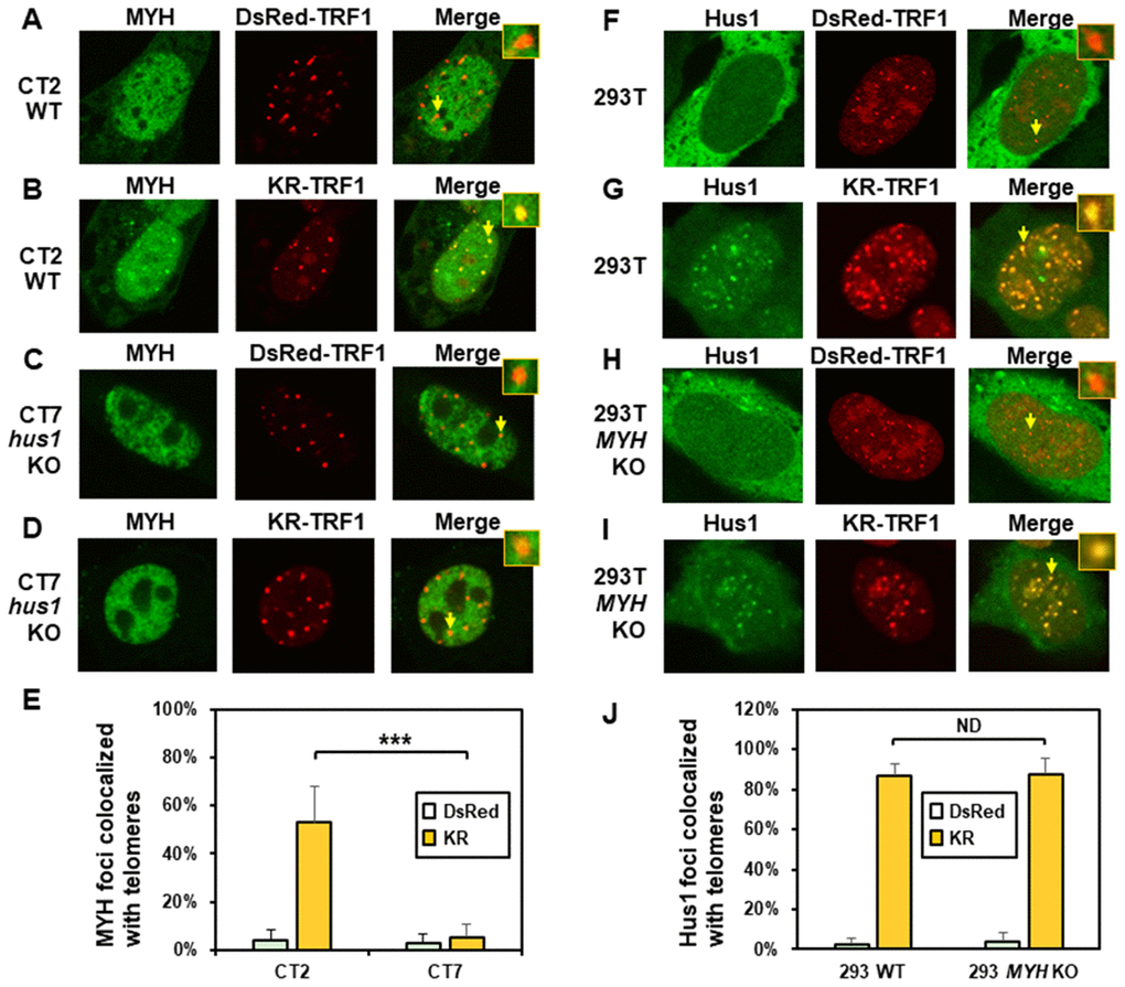
Figure 2.The formation of MYH foci induced at oxidatively damaged telomeres is dependent on Hus1. (A and C) GFP-MYH does not form foci at sites with DsRed-TRF1 in undamaged CT2 (hus1+/+) and CT7 (hus1-/-p21-/-) MEF cells, respectively. These cells contain granulated GFP-MYH spots and some of spots were localized with telomeres. (B and D) Damage response of GFP-MYH to the sites of KR-TRF1 after light activation in CT2 and CT7 MEF cells, respectively. GFP-MYH foci are not found at the sites of KR-TRF1 in CT7 cells. (E), Analyses of about 20 cells in each (A–D) group indicated that approximately 50% of GFP-MYH foci colocalized with telomeres in CT2 cells, in contrast, less than 5% of GFP-MYH foci colocalized with telomeres in CT7 cells. (F–J), GFP-Hus1 foci formation at telomeres in control HEK-293T and MYH KO HEK-293T human cells. Experiments were performed similarly to (A–E) except using GFP-hHus1 and different cells. After ROS induction by activating the KR protein, over 80% of GFP-Hus1 foci are colocalized with KR-TRF1 in both HEK-293T and MYH KO HEK-293T cells (J). ND indicates no difference.