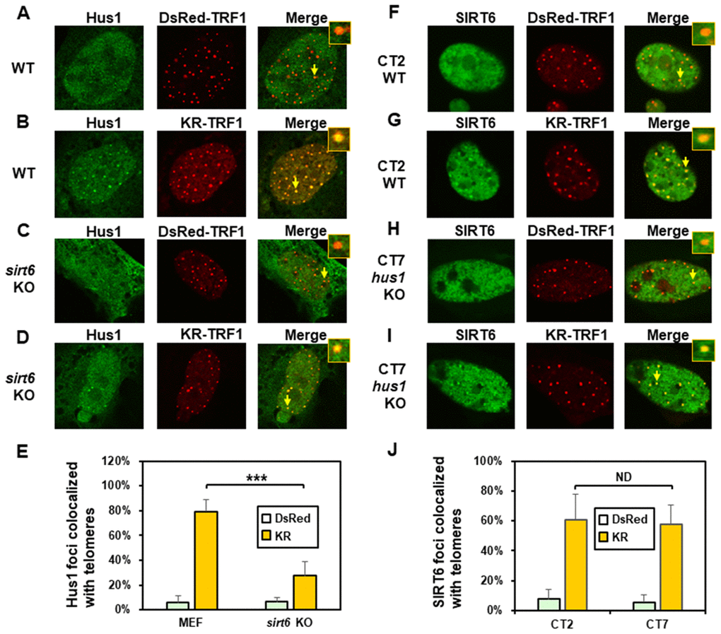
Figure 4.The formation of Hus1 foci induced at oxidatively damaged telomeres is partially dependent on SIRT6. (A and C), GFP-Hus1 does not form foci at sites with DsRed-TRF1 in undamaged control MEF and sirt6 KO MEF cells, respectively. (B and D), Damage response of GFP-Hus1 to the sites of KR-TRF1 after light activation in control and sirt6 KO MEF cells, respectively. GFP-hHus1 foci are significantly reduced at the sites of KR-TRF1 in sirt6 KO cells. (E), Analyses of about 20 cells in each (A–D) group indicated that approximately 80% of GFP-Hus1 foci colocalized with telomeres in control cells, in contrast, only 30% of GFP-Hus1 foci colocalized with telomeres in sirt6 KO cells. (F–J), GFP-hSIRT6 foci formation at telomeres in control CT2 and Hus1 KO CT7 MEF cells. Experiments were performed similarly to (A–E) except using GFP-hSIRT6 and different cells. After ROS induction by activating the KR protein, about 60% of GFP-SIRT6 foci are colocalized with KR-TRF1 in both CT2 and CT7 cells (J).