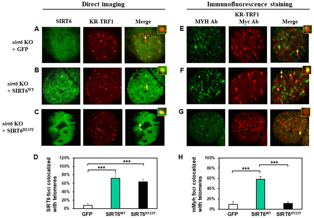
Figure 5.The recruitment of MYH at oxidatively damaged telomeres is dependent on the catalytic activities of SIRT6. MEF sirt6 KO cells were transfected with pEGFP-C1 vector or vector containing human wild-type (WT) or H133Y mutant hSIRT6 gene along with KR-TRF1 plasmid. (A–D), Both GFP-tagged hSIRT6WT and hSIRT6H133Y respond similarly to damaged telomeres. GFP-SIRT6 and Myc-tagged KR-TRF1 were detected by direct imaging. Images were captured 30 min after light activation with an Olympus FV1000 confocal microscopy system. (E–H), Response of endogenous mMyh to the damage sites in sirt6 KO cells expressing hSIRT6WT or hSIRT6H133Y mutant. Cell fluorescence was stripped by HCl treatment. GFP-SIRT6, MYH, and Myc-KR-TRF1 were them detected by immunofluorescence staining with GFP, MYH, and Myc antibodies (Ab), respectively, and reacted with blue, green, and red emission secondary antibodies, respectively. Cells containing blue-colored GFP-SIRT6 were selected for analyses to detect mMyh green foci and red-colored telomeres. (D) and (H), Quantitative analyses of 20 cells as in (A–C) and (E–F) groups, respectively. White, green, and black bars represent protein colocalization with telomeres in MEF sirt6 KO cells containing GFP alone, GFP-hSIRT6WT, or GFP-hSIRT6H133Y, respectively.