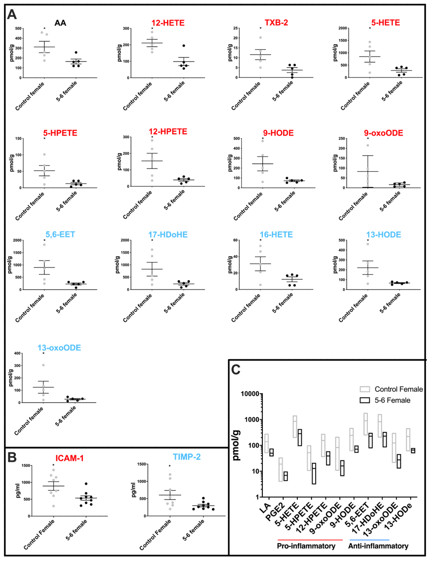
Figure 2.Molecular profiling of inflammation in female PD cerebellar mitochondria. (A) Oxylipin levels are lower in cerebellar mitochondria of PD Braak 5-6 females. Twelve oxylipin species and arachidonic acid are significantly reduced in the cerebellar mitochondria of females with PD Braak 5-6 when compared with controls. All samples were age matched. Braak 5-6 female n=5; control female n=5. (B) Two inflammatory cytokines are significantly lower in cerebellar mitochondria of PD Braak 5-6 females. ICAM-1 and TIMP-2 were significantly reduced in cerebellar mitochondria of PD Braak 5-6 females compared with female controls. Braak 5-6 female n=9; control female n=8. Plots display mean levels ± SEM. Red/blue titles represent pro- /anti-inflammatory, respectively. Refer to Supplementary Tables 3 and 5 for p values of oxylipins and inflammatory cytokines, respectively (Mann-Whitney U-test). (C) There is more variation in oxylipin content from cerebellar mitochondria of the female control group than the PD Braak 5-6 group. The female control group had significantly greater variance in eleven oxylipin quantities than PD Braak 5-6 females. PD Braak 5-6 female n=5; control female n=5. Box plots display interleaved high and low. The horizontal line represents the mean. Refer to Supplementary Table 4 for f values.
Figure 2 — Sex specific inflammatory profiles of cerebellar mitochondria are attenuated in Parkinson’s disease | Aging