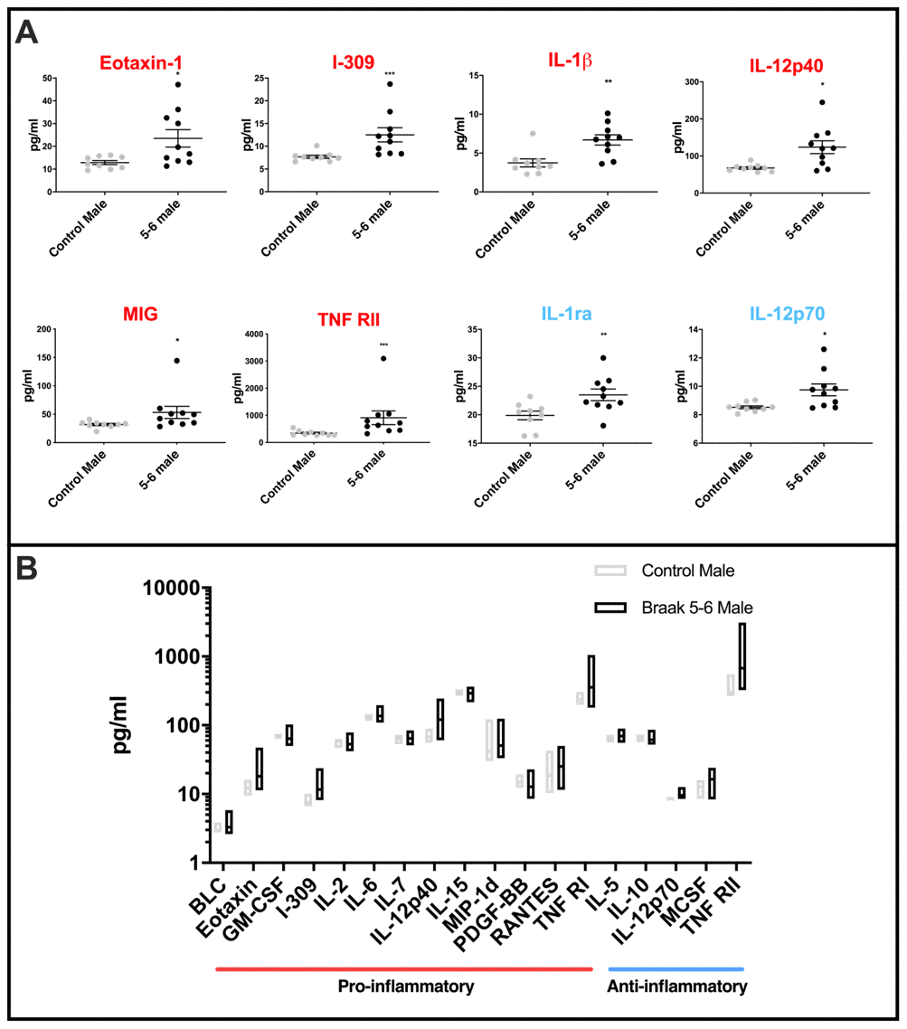
Figure 4.Cytokine levels in PD Braak 5-6 male cerebellar mitochondria (A) Mean levels of cytokines are higher in cerebellar mitochondria of PD Braak 5-6 males. Eight inflammatory cytokines were increased in cerebellar mitochondria of PD Braak 5-6 males compared with control males. PD Braak 5-6 male n=10; control male n=9. Red or blue titles represent pro- /anti-inflammatory, respectively. Plots display mean concentration (pg/ml) ± SEM (Mann-Whitney test), see Supplementary Table 5 for p values (Mann-Whitney U test). (B) PD Braak 5-6 males show different variance in cerebellar mitochondrial cytokines levels than was seen in the control male group. All seventeen measured inflammatory cytokines with significant differences in variance are more dispersed from the mean in PD Braak 5-6 males than control males. PD Braak 5-6 male n=10; PD Braak 5-6 female n=9; control male n=9; control female n=8. Box plots display interleaved high and low. The horizontal line represents the mean. Data shown are oxylipins (A+B) with significant variances (f-test). Refer to Supplementary Table 6 for f values.