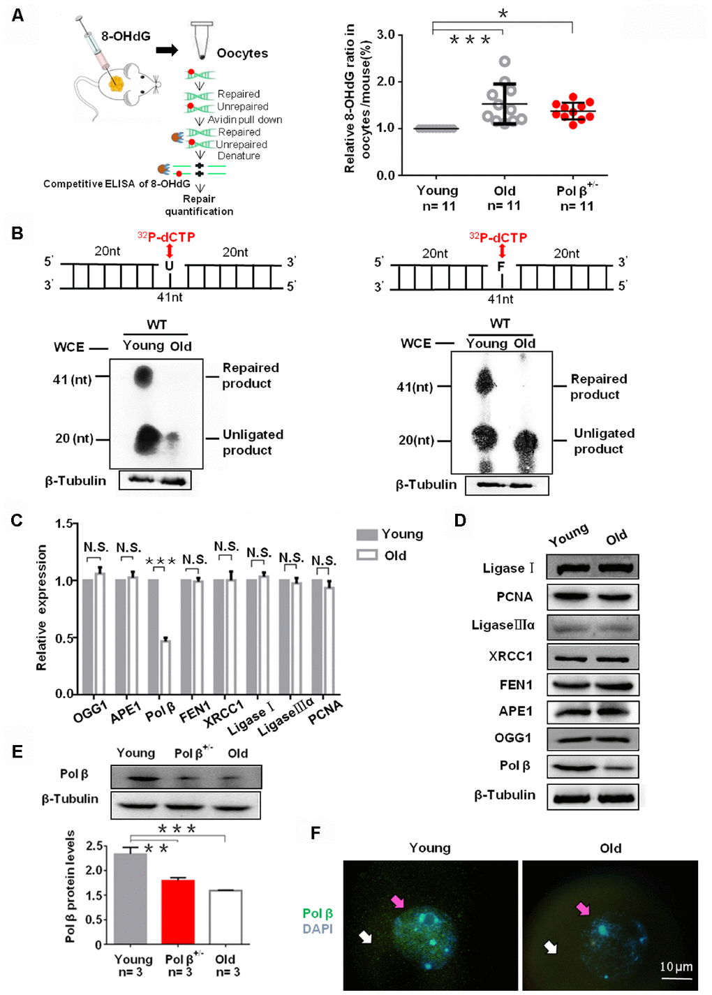
Figure 2.Aging reduce BER efficiency by low expression of Pol β in mouse oocytes. (A) Three equal dose of DNA oligo containing the damaged DNA lesion 8-OHdG was respectively transfected into young/old Pol β+/- mice ovary; 4h later, oocytes were lysed, and released 8-OHdG was determined by ELISA. With increasing age, reduced 8-OHdG repair was detected, indicating that age inhibits endogenous BER. Scatter graphs represent the content of 8-OHdG in oocytes, with a significant increase in the content of 8-OHdG in old (8 months) / Pol β+/- mice (6-8 weeks) compared to that in young mice (6-8 weeks) (n=11 per group; n stands for the number of mice, ***P<0.001, *P<0.05). (B) young/old oocytes, which were respectively divided into two parts for western blotting and BER assays, are defective in BER repair efficiency using whole-cell extract. The number of total oocytes in each line of the gel was detected to be approximately 500 and approximately 700, respectively. SP-BER: reconstitution with young and old oocytes. LP-BER: reconstitution with young and old oocytes. In this assay, whole young and old oocyte extracts were prepared to test SP-BER and LP-BER. Uracil or THF lesions were efficiently repaired by young oocytes but not by old oocytes. (C) Significant decrease in the expression of DNA repair genes in old mice (8 months) compared to that in young mice (6 to 8 weeks) shown by qRT-PCR. All results are the mean ± SD (n = 4 per group). Bar graphs represent the gene expression levels. The bar graphs show significantly lower levels of expression for Pol β in old mice than in young mice (***P < 0.001, Student’s t test). (D) Significant decrease in the expression of Pol β in old mice (8 months) compared to that in young mice (6 to 8 weeks) shown by western blotting, whereas, no significant difference in other genes. (E) Significant decrease in the expression of Pol β in old (8 months) / Pol β+/- mice (6-8 weeks) compared to that in young mice (6 to 8 weeks) shown by western blotting. We also show the quantitation with error bars (n=3 per group; n stands for the number of mice, *** P<0.001, **P<0.01). (F) Significant decrease in the expression of Pol β in old mice (8 months) compared to that in young mice (6 to 8 weeks) shown by photomicrographs. Representative photomicrographs show lower amounts of Pol β (green) protein expression in old mice than in young mice. Oocytes were counterstained with DAPI (blue). White arrows point to the cytoplasm and pink arrows to the nucleus. All bargraphs show the means ± SD.