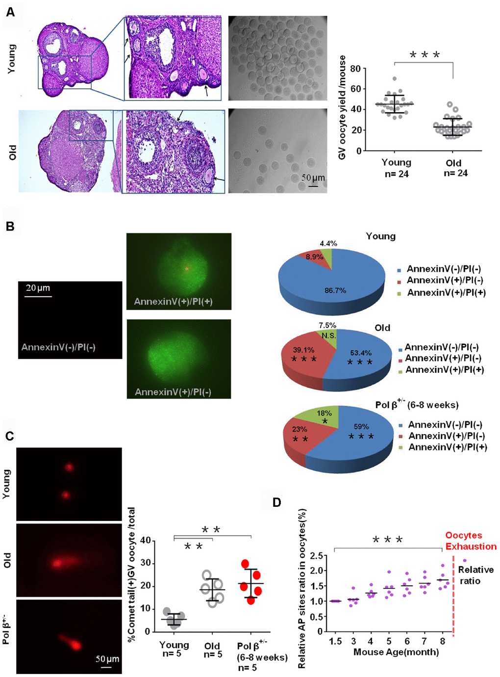
Figure 3.Aging-related ovarian reserve decline and SSBs from young and old mice. (A) Photomicrographs represent hematoxylin-eosin staining of young (upper) and old (below) C57BL/6J mice ovarian sections. Inset (upper left corner of young mice ovarian tissue) shows a higher number of oocytes yield; scatter graphs show a higher number of oocytes yield in young (6 to 8 weeks) compared to old (8 months) mice, (n =24 per group; n stands for the number of mice, ***P < 0.001, Student’s t test. The experiment was repeated six times). The black arrow indicates oocytes in ovarian follicles. (B) Immunofluorescence graphs show a result of Annexin V/PI staining. Statistics of oocytes number per young/ old/ Pol β+/- mouse including Annexin V(-)/PI(-), AnnexinV(+)/PI(+) and Annexin V(+)/PI(-). Adjacent pie charts show old/ Pol β+/- group more Annexin V(+)/PI(-), more Annexin V(+)/PI(+) and less Annexin V(-)/PI(-) compared to young group (6 to 8 weeks), (n=3 per group; ***P<0.001, ** P<0.01, *P<0.05). (C) The alkaline comet experiment shows that old/ Pol β+/- oocytes have more percentage of positive comet tail. (n=5 per group; n stands for the number of mice, **P<0.01, **P<0.01, Student’s t test). (D) Scatter graphs represent the relative ratio of AP sites in oocytes, with a significant increasing tendency in the content of AP sites in every age group with the increase of age (n=5 per group; ***P<0.001, Student’s t test).