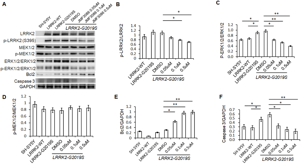
Figure 4.JMF3086 inhibited Lrrk2 MAPKKK activity and down-regulated ERK1/2 signaling pathways in SH-SY5Y cells stably expressing LRRK2-G2019S. (A) Western blot of total and phosphorylated target proteins, including Lrrk2, MEK1/2, ERK1/2, pro-apoptotic caspase 3, and anti-apoptotic Bcl2 after treatment with different concentrations of JMF3086. (B) Quantification of the effects of JMF3086 on the ratio of p-Lrrk2 to total Lrrk2. The relative expression of p-Lrrk2 to total Lrrk2 in LRRK2-WT and LRRK2-G2019S after treatment with DMSO solvent, 0.05 μM, 0.1 μM, and 0.5 μM JMF3086 was 0.92±0.08, 1.13±0.09, 1.09±0.08, 0.88±0.05, 0.78±0.03, and 0.70±0.04, respectively. P=0.09 for LRRK2-WT vs. LRRK2-G2019S; P=0.07 for LRRK2-G2019S with DMSO solvent vs. LRRK2-G2019S with 0.05 μM JMF3086; P=0.04 for LRRK2-G2019S with DMSO solvent vs. LRRK2-G2019S with 0.1 μM JMF3086; P=0.02 for LRRK2-G2019S with DMSO solvent vs. LRRK2-G2019S with 0.5 μM JMF3086, all one-way ANOVA. (C) Quantification of the effects of JMF3086 on the ratio of p-ERK1/2 to total ERK1/2. The relative expression of p-ERK1/2 to total ERK1/2 in SH-SY5Y controls, LRRK2-WT, and LRRK2-G2019S after treatment with DMSO solvent, 0.05 μM, 0.1 μM, and 0.5 μM JMF3086 was 0.63±0.05, 0.67±0.06, 0.92±0.10, 0.96±0.11, 0.63±0.09, 0.53±0.07, and 0.39±0.05, respectively. P=0.04 for SH-SY5Y controls or LRRK2-WT vs. LRRK2-G2019S; P=0.02 for LRRK2-G2019S with DMSO solvent vs. LRRK2-G2019S with 0.05 μM JMF3086; P=0.009 for LRRK2-G2019S with DMSO solvent vs. LRRK2-G2019S with 0.1 μM JMF3086; P=0.007 for LRRK2-G2019S with DMSO solvent vs. LRRK2-G2019S with 0.5 μM JMF3086, all one-way ANOVA. (D) Quantification of the effects of JMF3086 on the ratio of p-MEK1/2 to total MEK1/2. The relative expression of p-MEK1/2 to total MEK1/2 in SH-SY5Y controls, LRRK2-WT, and LRRK2-G2019S after treatment with DMSO solvent, 0.05 μM, 0.1 μM, and 0.5 μM JMF3086 was 0.95±0.08, 0.81±0.07, 0.78±0.08, 0.77±0.08, 0.86±0.05, 0.82±0.08, and 0.85±0.08, respectively. (E) Quantification of the effects of JMF3086 on the ratio of Bcl2 to GAPDH. The relative expression of Bcl2 to GAPDH in SH-SY5Y controls, LRRK2-WT, and LRRK2-G2019S after treatment with DMSO solvent, 0.05 μM, 0.1 μM, and 0.5 μM JMF3086 was 0.25±0.02, 0.07±0.01, 0.21±0.02, 0.30±0.01, 0.62±0.10, 0.95±0.12, and 0.98±0.14, respectively. P=0.02 for LRRK2-G2019S with DMSO solvent vs. LRRK2-G2019S with 0.05 μM JMF3086; P=0.009 for LRRK2-G2019S with DMSO solvent vs. LRRK2-G2019S with 0.1 μM JMF3086; P=0.008 for LRRK2-G2019S with DMSO solvent vs. LRRK2-G2019S with 0.5 μM JMF3086, all one-way ANOVA. (F) Quantification of the effects of JMF3086 on the ratio of caspase-3 to GAPDH. The relative expression of caspase-3 to GAPDH in SH-SY5Y controls, LRRK2-WT, and LRRK2-G2019S after treatment with DMSO solvent, 0.05 μM, 0.1 μM, and 0.5 μM JMF3086 was 0.30±0.07, 0.28±0.06, 0.47±0.08, 0.58±0.05, 0.32±0.07, 0.24±0.08, and 0.20±0.08, respectively. P=0.04 for SH-SY5Y controls or LRRK2-WT vs. LRRK2-G2019S; P=0.03 for LRRK2-G2019S with DMSO solvent vs. LRRK2-G2019S with 0.05 μM JMF3086; P=0.02 for LRRK2-G2019S with DMSO solvent vs. LRRK2-G2019S with 0.1 μM JMF3086; P=0.009 for LRRK2-G2019S with DMSO solvent vs. LRRK2-G2019S with 0.5 μM JMF3086, all one-way ANOVA. All neurons were treated for 48 h and then lysed. Equal amounts of protein lysate were subjected to SDS-PAGE and the proteins analyzed by Western blotting. Immunoblots were probed with the indicated antibodies. All experiments were repeated three times. Data represent mean ± SEM. *P<0.05, **P<0.01.