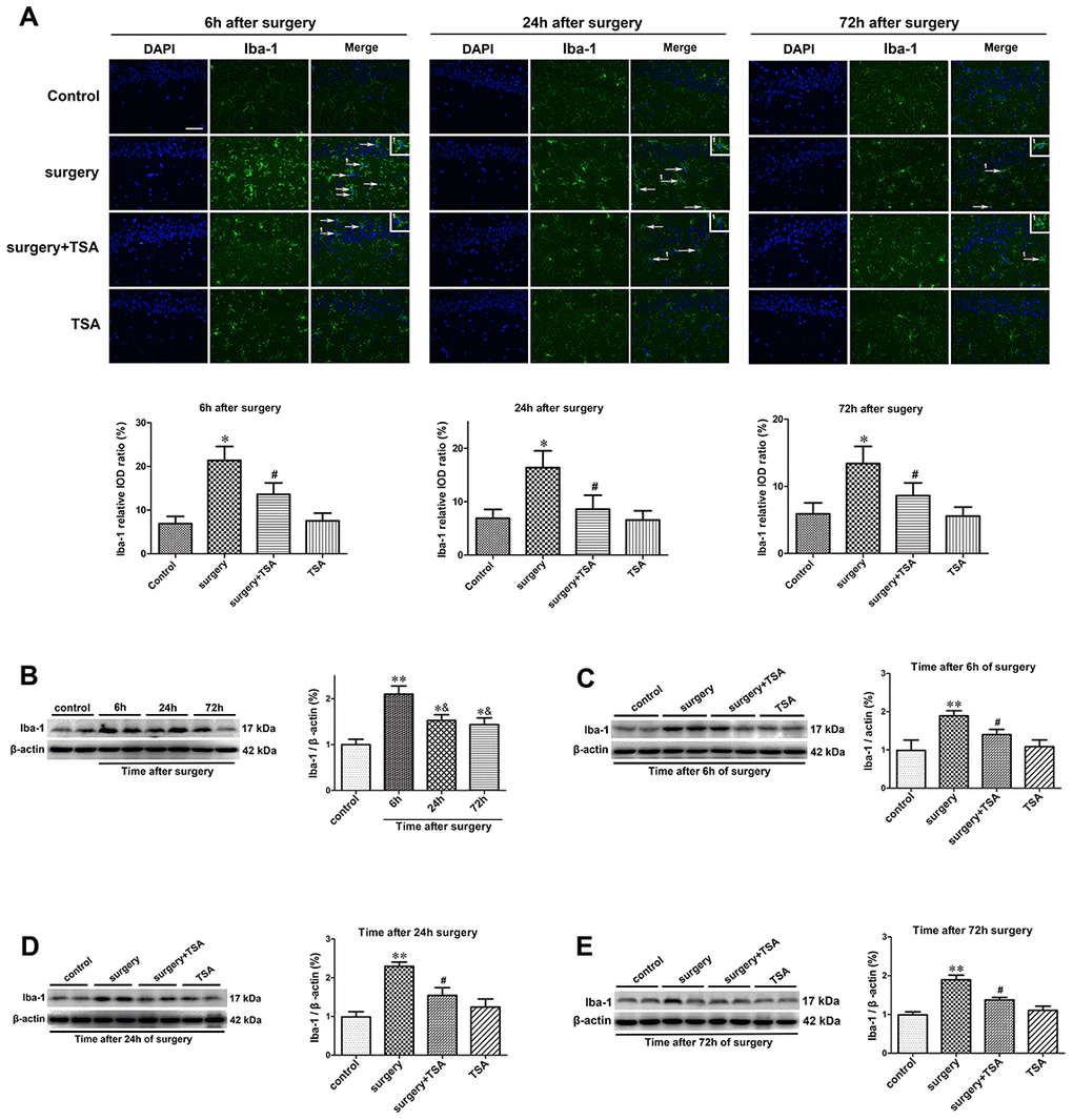
Figure 2.TSA inhibits surgery-induced hippocampal microglia activation in aged rats. (A) Immunofluorescence analysis and semi-quantification of higher expression of Iba-1 protein in the hippocampal CA1 area were observed at postoperative 6 h, 24 h and 72 h, and this staining was significantly inhibited by TSA pretreatment (Iba-1, green; cell nuclei, blue. Magnification 400 ×, Scale bar = 50 μm). Western blot analysis and semi-quantitative data showing protein expression (B–E), indicating that the expression levels of Iba-1, a microglia activation marker, increased significantly at 6 h, 24 h and 72 h after laparotomy, and it peaked at 6 h and decreased within 72 h post-surgery, which was significantly inhibited by TSA pretreatment, with β-actin used as a loading control. Data are given as means {plus minus} SEM, n = 5. *P < 0.05 and **P < 0.01 vs. the control group; #P < 0.05 vs. the surgery group; &P < 0.05 vs. 6 h after surgery.