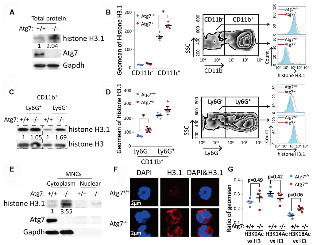
Figure 3.Atg7-deletion accumulated histone H3.1 protein with incorrect cytoplasmic localization in the bone marrow CD11b+Ly6G- myeloid cells. (A, C). Western blotting analysis of histone H3.1 in bone marrow cells. Gapdh or total histone H3 was used as a loading control. (A) Mononuclear cells; (C) CD11b+Ly6G- and CD11b+Ly6G+ myeloid cells. (B, D) Flow cytometric analysis of protein level of histone H3.1 in bone marrow cells. (B) Analysis of histone H3.1 in CD11b- and CD11b+ bone marrow cells. (D) Analysis of histone H3.1 in CD11b+Ly6G- and CD11b+Ly6G+ myeloid cells. Right, scheme for analysis of histone H3.1 in bone marrow cells. Left, statistical analysis of histone H3.1 geometric mean fluorescence intensity (MFI) in bone marrow cells. (E) Western blotting analysis of histone H3.1 in cytoplasm and nucleus from mononuclear cells. (F). Confocal detection of histone H3.1 protein in CD11b+Ly6G- myeloid cells. (G) Ratio of geometric mean of H3K9/14/18Ac compared to H3.