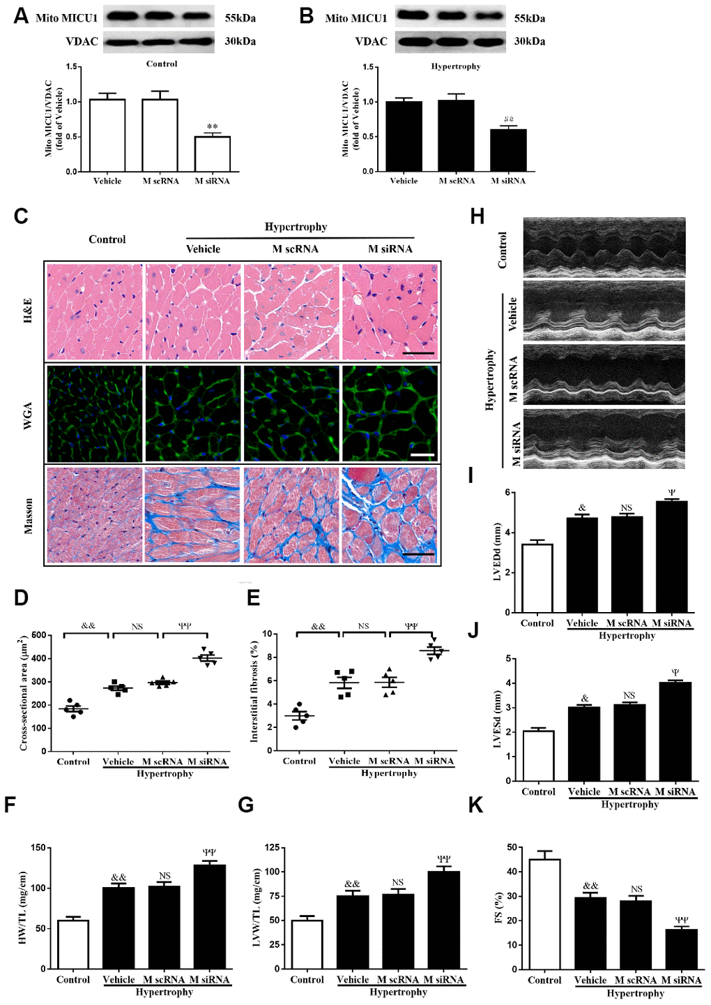
Figure 2.MICU1 downregulation in the heart aggravated Ang-II-induced cardiac hypertrophy. (A) Western blotting was used to measure the transfection efficiency of MICU1 siRNA in control mice. (B) Transfection efficiency of MICU1 siRNA in mice subjected to Ang-II was determined by Western blotting. (C) The heart sections stained with hematoxylin-eosin (H&E, the first row; Scale bars=50 μm), wheat germ agglutinin (WGA, the second row; Scale bars=20 μm) and Masson (the third row; Scale bars=50 μm) from the indicated groups were measured. (D) Cross-sectional cardiomyocyte areas were summarized. (E) The interstitial fibrosis was quantified. (F, G) The ratio of heart weight to tibia length (HW/TL) (F) and left ventricular weight to tibia length (LVW/TL) (G) were determined in different mice. (H) Representative echocardiographic image of the left ventricle in different mice was represented. (I–K) Echocardiographic assessment of left ventricular end-diastolic dimension (LVEDd) (I), left ventricular end-systolic dimension (LVESd) (J) and fractional shortening (FS) (K) was used to reflect cardiac function. M scRNA, scrambled siRNA; M siRNA, MICU1-specific siRNA. All the data represent the means ± SEM. N=6-8/group. **P<0.01 vs. Vehicle in Control; ##P<0.01 vs. Vehicle in Hypertrophy; &P<0.05, &&P<0.01 vs. Control; ΨP<0.05, ΨΨP<0.01 vs. M scRNA of hypertrophy.