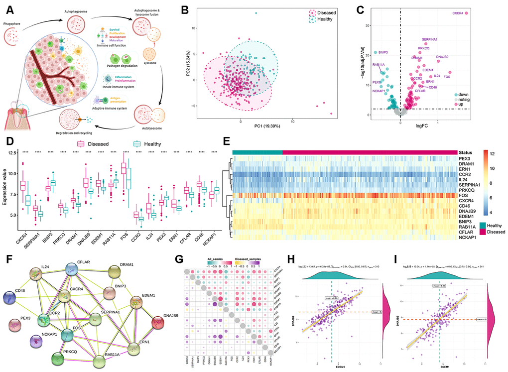
Figure 1.Expression landscape autophagy genes in periodontitis. (A) The overview of autophagy in regulating dynamic homeostasis of the immune microenvironment in periodontitis. (B) Principal component analysis (PCA) of 208 autophagy genes between healthy and periodontitis. The two first principal components (PC1, PC2) which could explain most of the variables are plotted, suggesting there are diversity regulation patterns of autophagy between healthy and periodontitis. (C) The volcano-plot shows the summary of expression changes of 208 autophagy genes between healthy and periodontitis samples and the most significant 16 autophagy genes are labeled. (D, E) The box-plot and heatmap-plot demonstrated the transcriptome expression status of 16 significantly dysregulated autophagy genes between healthy and periodontitis samples. (F) The 16 significant dysregulated autophagy gene protein-protein interactions are presented. (G) Correlations among the expression of 16 significantly dysregulated autophagy genes in all samples and periodontitis samples. (H, I) The two scatter-plots demonstrated the most correlated two autophagy genes: EDEM1 and DNAJB9.