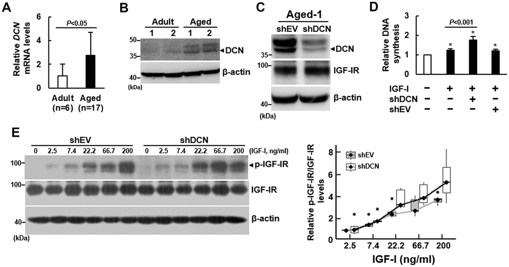
Figure 3.Effect of DCN knockdown on the DNA synthesis and IGF-IR auto-phosphorylation of aged bmMSCs. (A) RT-qPCR analyses. The expression of DCN mRNA of bmMSCs from 17 aged donors was compared to that of the cells from 6 adult donors (to which a value of 1 was assigned). Data represent the mean ± S.D. from a triplicate analysis. Student’s t-test was used to analyze the differences between the groups. (B) Western blot analyses of DCN levels. Representative blots of the DCN levels in the Aged-1, Aged-2, Adult-1, and Adult-2 cells are shown. (C) Western blot analysis of DCN and IGF-IR levels in cells with DCN knockdown. Aged-1 cells were infected with Lenti virus to generate DCN-knockdown (chDCN) and empty vector control (shEV) cells. The DCN and IGF-IR protein levels of the parental Aged-1, shDCN, and shEV cells are shown. (D) BrdU incorporation analyses. Serum-starved parental, shDCN, and shEV cells were treated with 200 ng/ml IGF-I for 24h, and examined for the IGF-I-induced DNA synthesis. The DNA syntheses in these cells were compared to that of the untreated parental cells (to which a value of 1 was assigned). Data represent the mean ± S.D. from three experiments. A one-way ANOVA plus Scheffe’s post hoc tests were used to analyze the differences among the untreated and IGF-1-treated groups. *, P<0.05 versus untreated control. Student’s t-test was used to analyze the differences between the groups. (E) Western blot analyses of IGF-IR auto-phosphorylation. Serum-starved shDCN and shEV cells were either treated with varying doses of IGF-I for 5 min or left untreated. IGF-IR auto-phosphorylation was examined and normalized to total IGF-IR expression. The difference in the response rates between shDCN and shEV cells was analyzed by linear regression analyses.