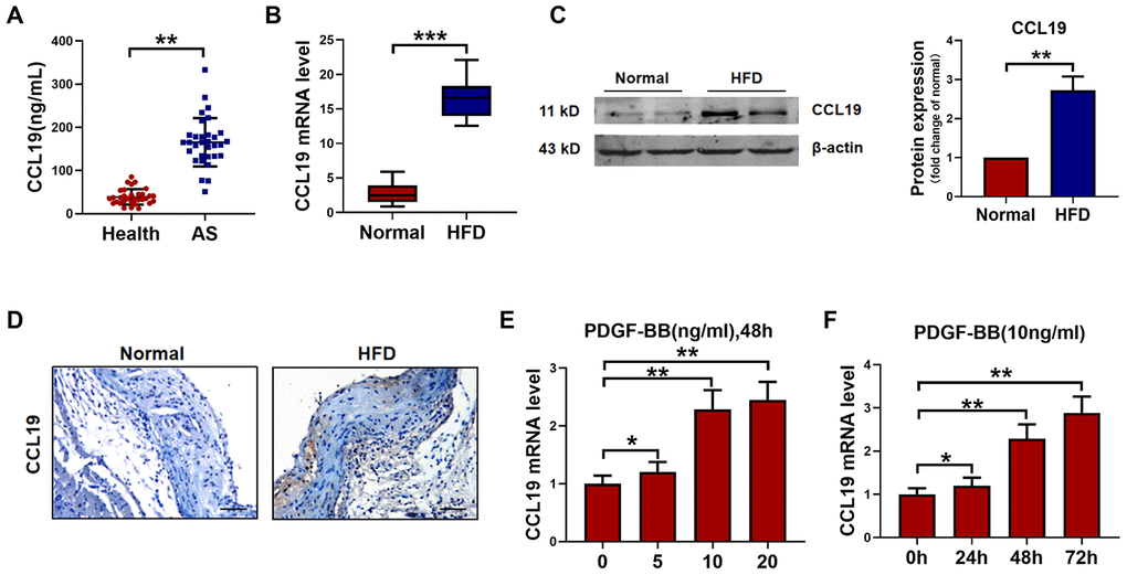
Figure 1.CCL19 expression in AS. (A) Detection of CCL19 expression in peripheral blood of patients by ELISA. n=32. (B) qRT-PCR analysis of CCL19 mRNA levels normalized to GAPDH in AS mice aorta and normal aorta tissues. n=10. (C) CCL19 protein levels in the AS or normal mice aorta tissues was performed by western blot. n=4. (D) Representative images of IHC of CCL19 in AS model mice aorta tissues and normal aorta tissues. Bar=50 μm. The effect of different concentrations (E) or different duration (F) of PDGF-BB on the expression of CCL19 was detected by qRT-PCR. n=5. *P<0.05; **P<0.01; ***P<0.001.